The Study of Tissues: Histology
Laboratory Exercise – Tissues and Histology
Tissues are made of similar groups of cells that work together to carry out a specific function. Humans are multicellular organisms; the cells in our tissues work in a coordinated and synchronized effort in conjunction with other types of tissues. The human body has four main categories of tissues: epithelial, connective, muscular and neural tissues.
The branch of biology that studies the microscopic structure of tissues is called histology.
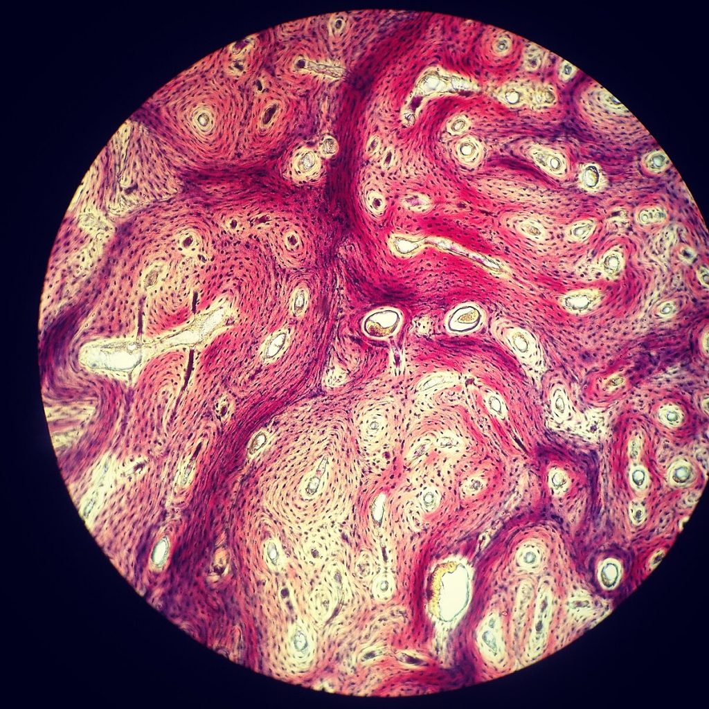
Learning Outcomes
- List the 4 main types and characteristics of tissue found in the human body
- Identify different types of epithelial tissue based on cell morphology and number of layers.
- List the location and function of selected epithelial tissues in the human body
- Identify different types of connective tissue.
- List the location and function of selected connective tissues in the human body
- Identify the three types of muscle tissue found in the human body.
- List the location and function of three types of muscle tissue in the human body
- Identify cell types in nervous tissue.
- Describe the function of cells found in nervous tissue.
Laboratory supplies
Compound microscopes
Prepared slides:
Kidney – capsule of Bowman’s – simple squamous epithelium
Lung – alveoli – simple squamous epithelium
Kidney – tubules x.s. and l.s – simple cuboidal epithelium
Small intestine – simple columnar epithelium
Esophagus – non keratinized stratified squamous epithelium
Thick Skin – keratinized stratified squamous epithelium
Thin skin – sweat glands – stratified cuboidal epithelium
Trachea – pseudostratified columnar ciliated epithelium
Urinary bladder – transitional epithelium
Areolar CT –
Adipose CT –
Reticular CT – lymph node
Dense regular connective tissue – Tendon
Dense irregular connective tissue – skin (dermis)
Elastic connective tissue – Aorta
Hyaline cartilage
Elastic cartilage
Fibrocartilage
Compact bone – ground
Human blood smear
Cardiac muscle – intercalated disk stain
Smooth muscle
Skeletal muscle
Neuronal smear
Notebook, pencil, eraser, color pencils
Pre-Laboratory Activities – Tissues and Histology Laboratory
It is expected that students read the corresponding chapter of the assigned textbook (Tissues), view videos/animations (if applicable) and then answer the questions shown below before coming to the laboratory.
Epithelial Tissue
View “Classification of Epithelia – Drawn & Defined” video from YouTube using the link shown below
Complete the following questions
- Draw a typical epithelial cell (at least two of them together) and label the apical portion, microvilli, nucleus, tight junctions, desmosomes, hemidesmosomes and basal portion.

- List the three basic epithelial cell shapes

- How is epithelial tissue classified?

- Name the two types of epithelia that are not classified based on the characteristic you mentioned above?

- Fill in the table shown below; epithelial tissue and its location. You will use it as reference during the lab activity.
| Type of epithelial tissue | Locations |
| Simple squamous epithelium | |
| Simple cuboidal epithelium | |
| Simple columnar epithelium | |
| Pseudostratified columnar ciliated epithelium with goblet cells | |
| Stratified squamous epithelium (non-keratinized) | |
| Stratified squamous epithelium (keratinized) | |
| Stratified cuboidal epithelium | |
| Stratified columnar epithelium | |
| Transitional epithelium |
Connective Tissue
View “Tissues, Part 4 – Types of Connective Tissues: Crash Course Anatomy & Physiology #5” video from YouTube using the link shown below:
Answer the following questions:
- List the three shared characteristics of connective tissue.

- Name 3 types of fibers found in connective tissue.

- Define the following terms:
- Fibroblast

- Chondrocyte

- Osteocyte

- Matrix

- Fibroblast
Muscular Tissue
View “Three types of muscle – Khan Academy” video from YouTube using the link shown below
Answer the following questions
- List the three types of muscular tissue

- List the characteristics of skeletal muscle

- List the characteristics of smooth muscle

- List the characteristics of cardiac muscle

Nervous Tissue
View the following videos
- “Introduction to neural cell types – Khan academy” video from YouTube using the link shown below:
- “The neuron” video from YouTube using the link shown below:
Answer the following questions
- Name the two types of cells present in nervous tissue. Describe their function.

- Draw and label a typical motor neuron (include: dendrites, body, mitochondria, endoplasmic reticulum system, nucleus, axon, Schwan cells, telodendria)

Introduction to Tissues and Histology
Tissues are defined as collections of cells and the extracellular substances surrounding them that gather to perform a specific function. There are four main types of human tissue: epithelial, connective, muscular, and nervous. Histology is the microscopic study of tissues.
Epithelial Tissue
The epithelium consists of cells of diverse shapes that contain little extracellular matrix. The epithelium covers surfaces and lines the lumen of internal hollow organs, usually has an apical portion, a basement membrane for attachment, and it is avascular. The main function of epithelial tissue is to protect structures, it acts as barriers allowing only some substances to pass through epithelial layers. Epithelium also secretes and absorb substances depending on its location.
Epithelium is mostly classified based on the morphology of the cells and the number of cell layers present. The types of cells can be squamous (flat), cuboidal and columnar. Epithelium may be simple (one layer) or stratified (more than one layer of cells).
You can combine both cell shape and number of layers to have the following types of epithelium:
- Simple squamous
- Stratified squamous (nonkeratinized or keratinized)
- Simple cuboidal
- Stratified cuboidal
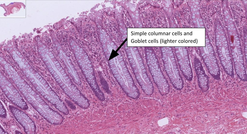
- Simple columnar
- Stratified columnar
Besides those we also have other two types of epithelia that are not classified based on cell shape and number of layers:
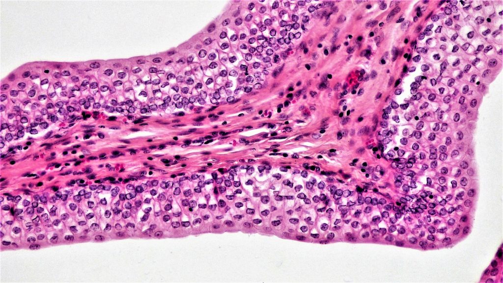
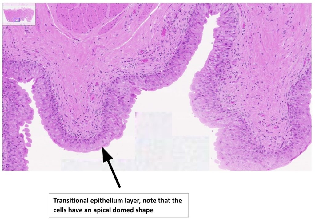
- Transitional epithelium (stratified, with cells that can change shape, cells have an apical dome shape),
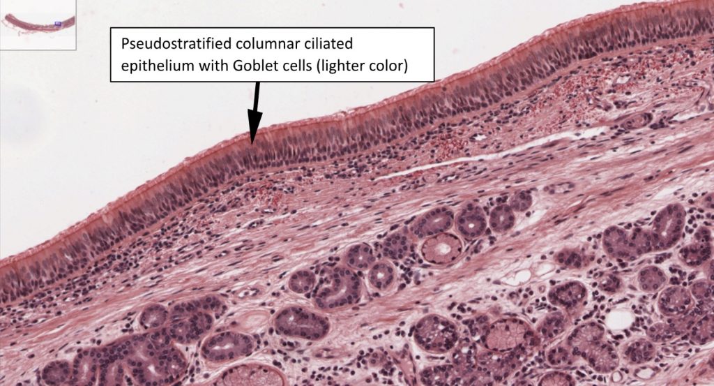
- Pseudostratified columnar ciliated epithelium – has a single layer of cells that appears stratified, because some cells do not reach the apical portion of the layer.
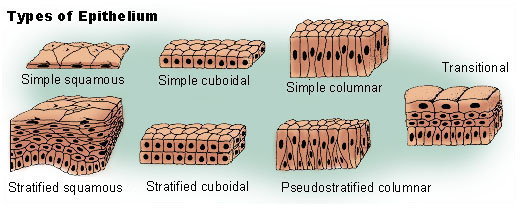
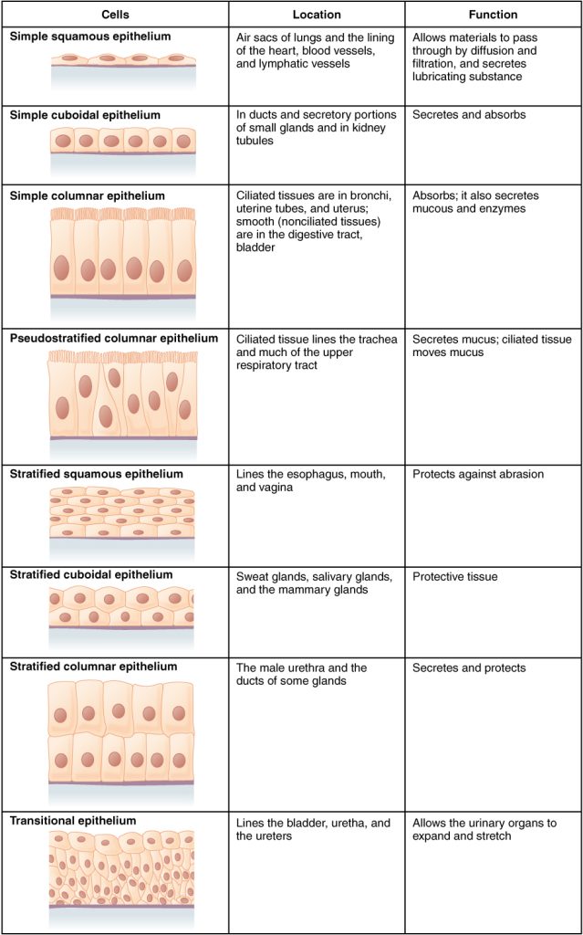
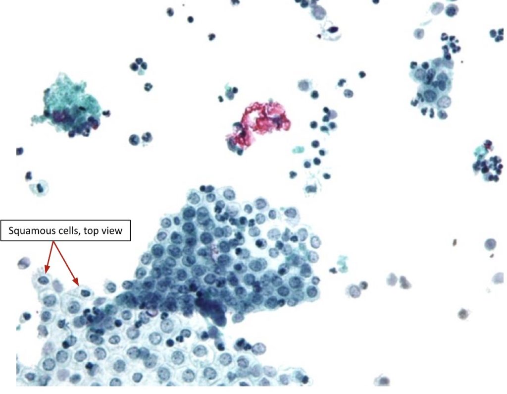
Selected histological images of Epithelial tissue
Virtual Microscope
Use the QR code shown below to access the Virtual Microscope from University of Michigan. Use the +/- signs below the image to increase/decrease magnification. You may also scan the image – look at the top left of the main view, you will see a smaller window with a blue square, drag the square to change the area where you like to see. You can also move to a different area in the main view by dragging the small white hand shown there.

Simple cuboidal epithelium
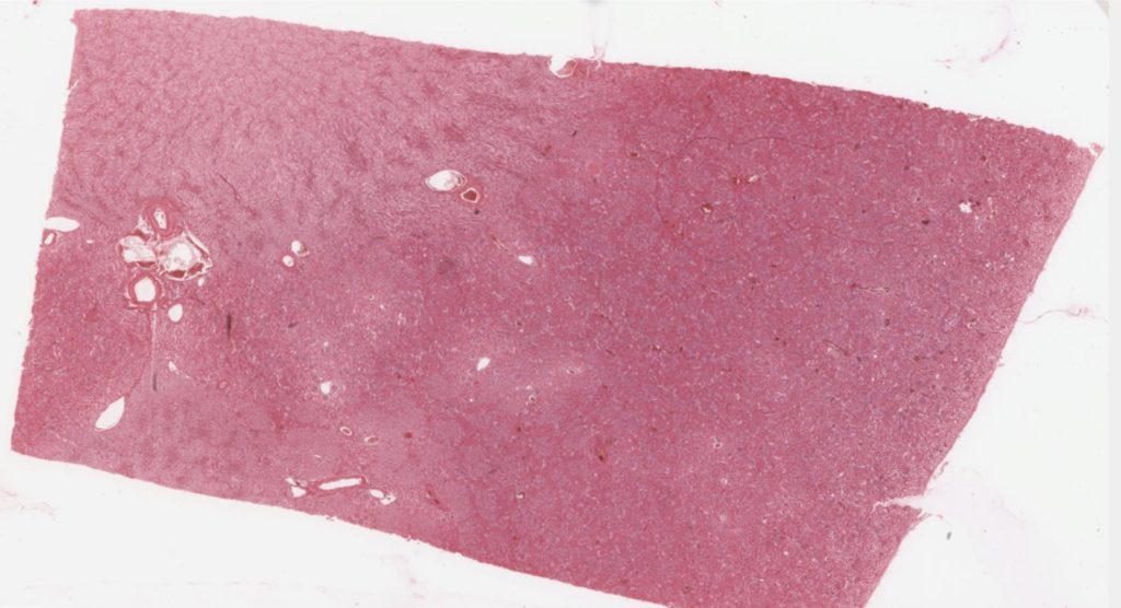
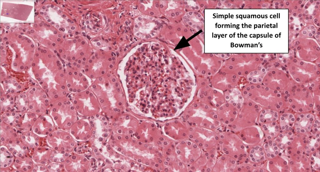
Then, focus on the area shown below (capsule of Bowman’s, parietal layer – containing simple squamous epithelium – flat cells:
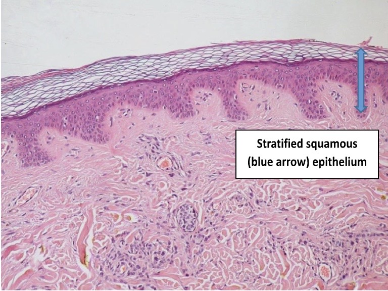
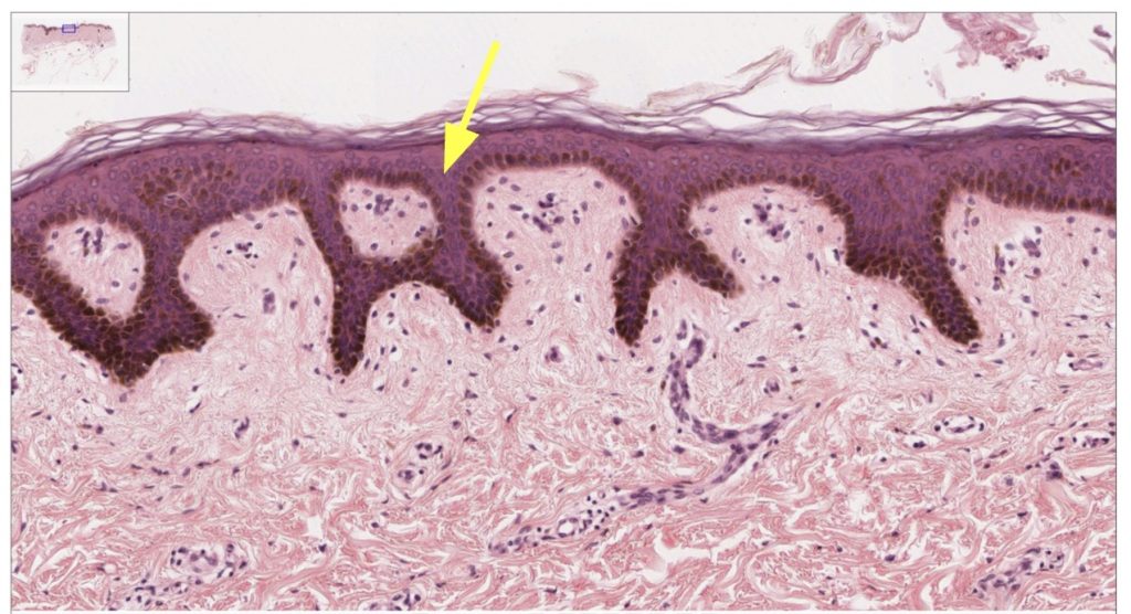
Stratified squamous epithelium (keratinized)
Virtual Microscope
Use the QR code shown below to access the Virtual Microscope from University of Michigan. Use the +/- signs below the image to increase/decrease magnification. You may also scan the image – look at the top left of the main view, you will see a smaller window with a blue square, drag the square to change the area where you like to see. You can also move to a different area in the main view by dragging the small white hand shown there.

Thin skin, H&E, 40X (104-2)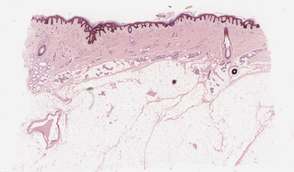
Then focus on the epidermal layer – contains stratified squamous epithelium
Simple cuboidal epithelium
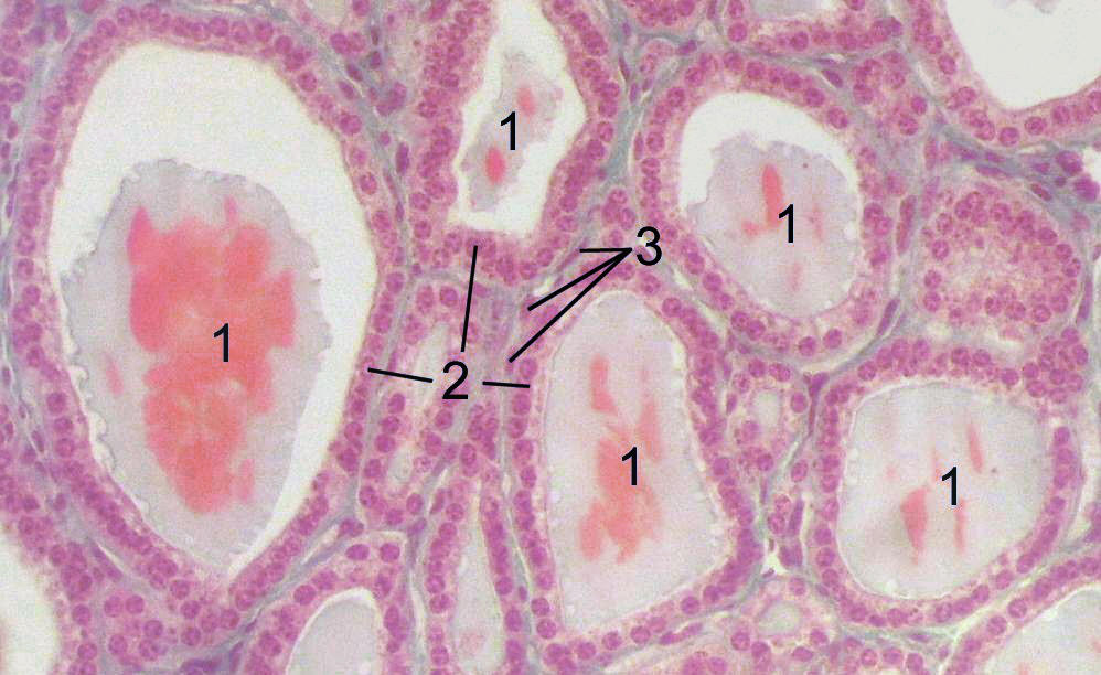
This is a microscope side of thyroid; showing follicles made of follicular cells (cuboidal shape), labeled #2 and filled with a colloid substance, labeled #1. The cells located between the follicles, labeled #3 are also of cuboidal shape and are called parafollicular cells.
Virtual Microscope – use the QR code shown below to access the Virtual Microscope from University of Michigan. Use the +/- signs below the image to increase/decrease magnification. You may also scan the image – look at the top left of the main view, you will see a smaller window with a blue square, drag the square to change the area where you like to see. You can also move to a different area in the main view by dragging the small white hand shown there.

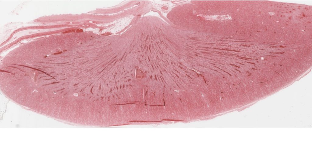
Kidney, human, H&E, 40X (cortex, glomerulus, proximal tubule, distal tubule, collecting duct, cortical labyrinth, medullary ray, medulla, vasa recta, loop of Henle, arcuate artery, interlobular arteries and veins). Cuboidal cells are located in the proximal tubule, distal tubule, collecting duct.
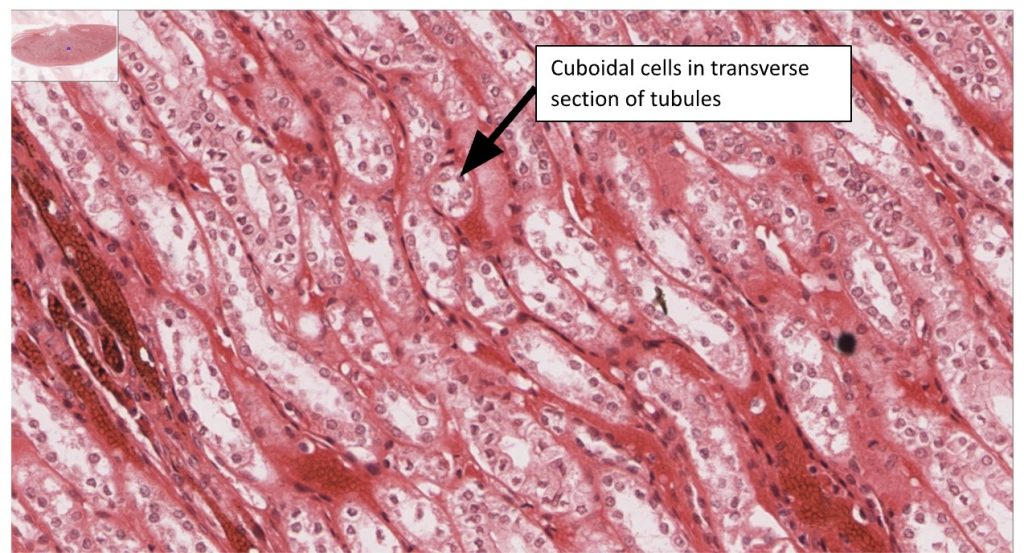
Then focus on the area where the tubules of the nephron are located to be able to see the simple cuboidal epithelium.
Simple columnar epithelium
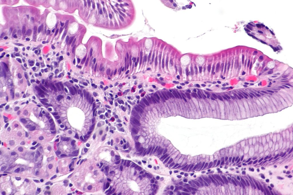
Virtual Microscope – use the QR code shown below to access the Virtual Microscope from University of Michigan. Use the +/- signs below the image to increase/decrease magnification. You may also scan the image – look at the top left of the main view, you will see a smaller window with a blue square, drag the square to change the area where you like to see. You can also move to a different area in the main view by dragging the small white hand shown there.

Colon, H&E, 40X (simple columnar epithelium) #176
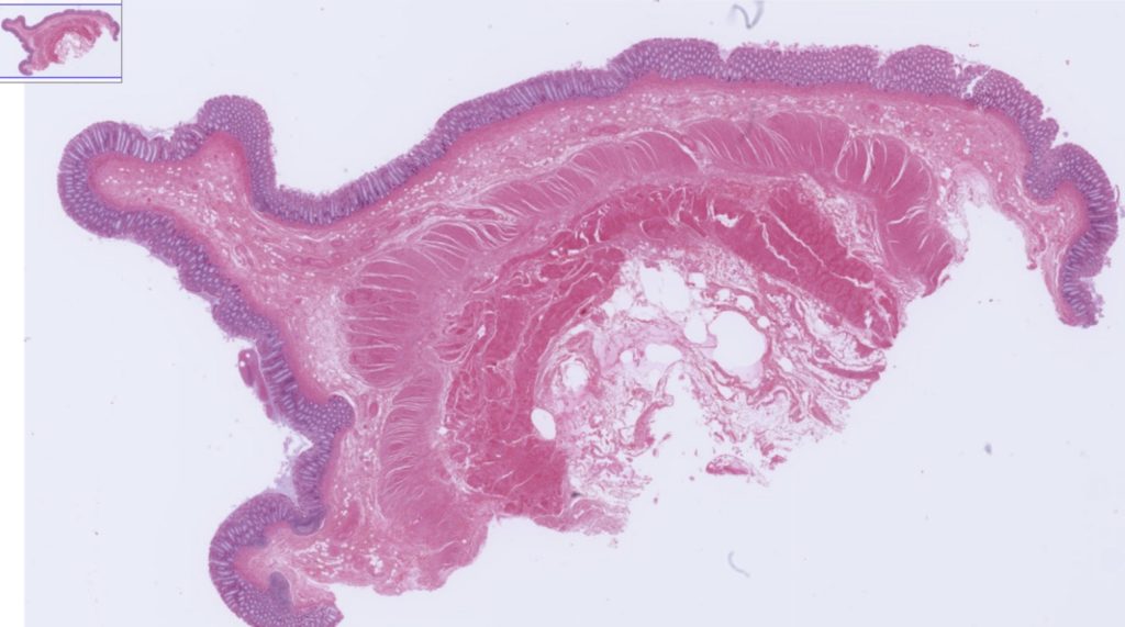
Then focus on the layer that contains simple columnar cells
Pseudostratified columnar ciliated epithelium
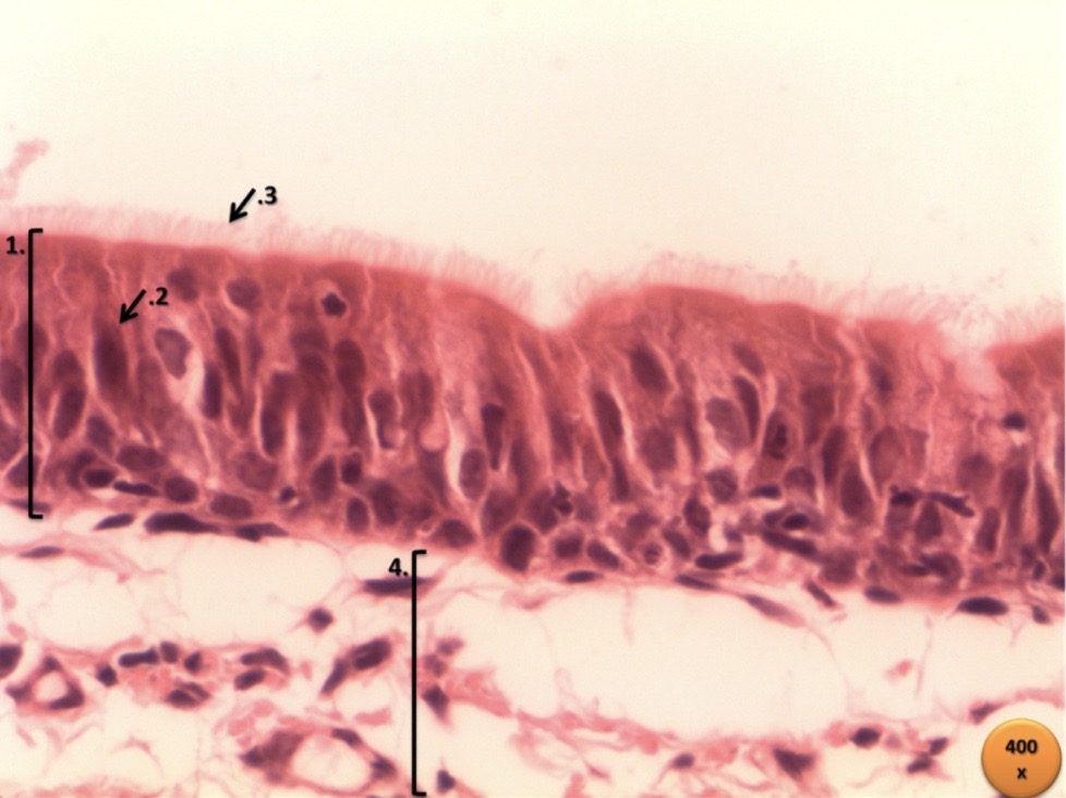
Virtual Microscope – use the QR code shown below to access the Virtual Microscope from University of Michigan. Use the +/- signs below the image to increase/decrease magnification. You may also scan then image – look at the top left of the main view, you will see a smaller window with a blue square, drag the square to change the area where you like to see. You can also move to a different area in the main view by dragging the small white hand shown there.

Trachea H&E, 40X (pseudostratified columnar epithelium), #040

Then focus on the layer that contains pseudostratified columnar ciliated epithelium.
Transitional Epithelium
Virtual Microscope – use the QR code shown below to access the Virtual Microscope from University of Michigan. Use the +/- signs below the image to increase/decrease magnification. You may also scan the image – look at the top left of the main view, you will see a smaller window with a blue square, drag the square to change the area where you like to see. You can also move to a different area in the main view by dragging the small white hand shown there.

Non-distended bladder, H&E, 40X (transitional epithelium), # Duke University 98.
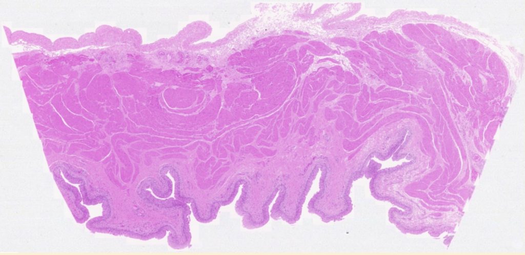
Then focus on the transitional epithelium layer
Connective Tissue
Connective tissues connect one tissue to one another; acts as support and participate in movement of body parts. It also stores compounds such as calcium and phosphorous; it cushions and insulate; and enclose organs. There are many types of connective tissue; they look very different, but they share their origin and 3 main characteristics; they contain ground substance, fibers (collagen, elastic, reticular) and specialized cells (adipose cells, chondrocytes, osteocytes, blood cells, macrophages, and mesenchymal cells cells). The combination of ground substance (Hyaluronic acid and proteoglycans) and fibers is also known as the Matrix.


Connective tissue classification is based on the type specialized cells and the extracellular matrix. There are two main types of connective tissue:
- Embryonic connective tissue is called mesenchyme, consists of irregularly shaped cells and abundant matrix, and gives rise to adult connective tissue.
- Adult connective tissue consists of connective tissue proper, supporting connective tissue, and fluid connective tissue. The adult connective tissue is further subclassified as
Connective Tissue Proper
- Loose connective tissue
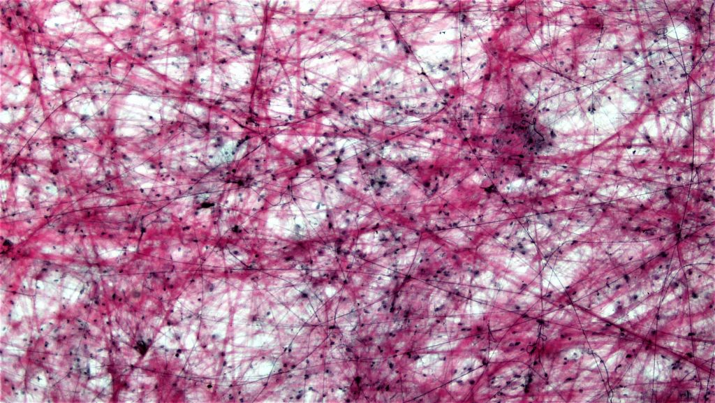
Loose Areolar connective tissue has many different cell types and a random arrangement of protein fibers with space between the fibers. This tissue fills spaces around the organs and attaches the skin to underlying tissues.
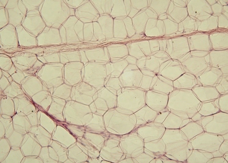
Adipose tissue has adipocytes filled with lipid and very little extracellular matrix. It protects body structures, also serves as a place for energy storage, and insulation.
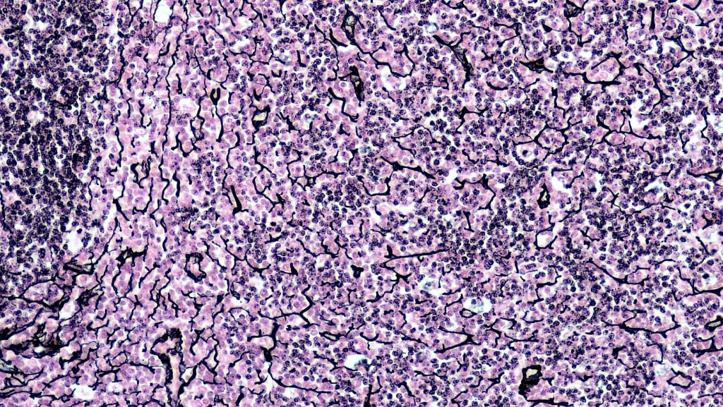
Reticular tissue is a network of reticular fibers, found in lymphatic tissue, bone marrow, and the liver.
b. Dense connective tissue
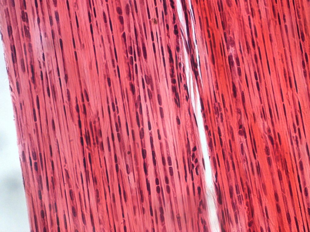
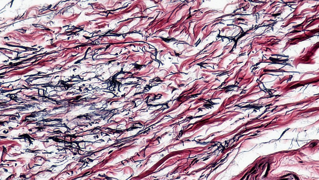
Dense regular connective tissue is composed of fibers regularly arranged in one direction. There are two types of dense regular connective tissue; collagenous (tendons and most ligaments) and elastic (ligaments of vertebrae).
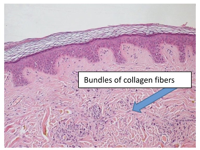
Dense irregular connective tissue has fibers organized in many directions, there are two types of dense irregular connective tissue: collagenous (found as capsules of organs and in the dermis of skin) and elastic (found in large arteries, such as the aorta).
Dense irregular connective tissue
Elastic connective tissue
Supporting Connective Tissue
-
Cartilage
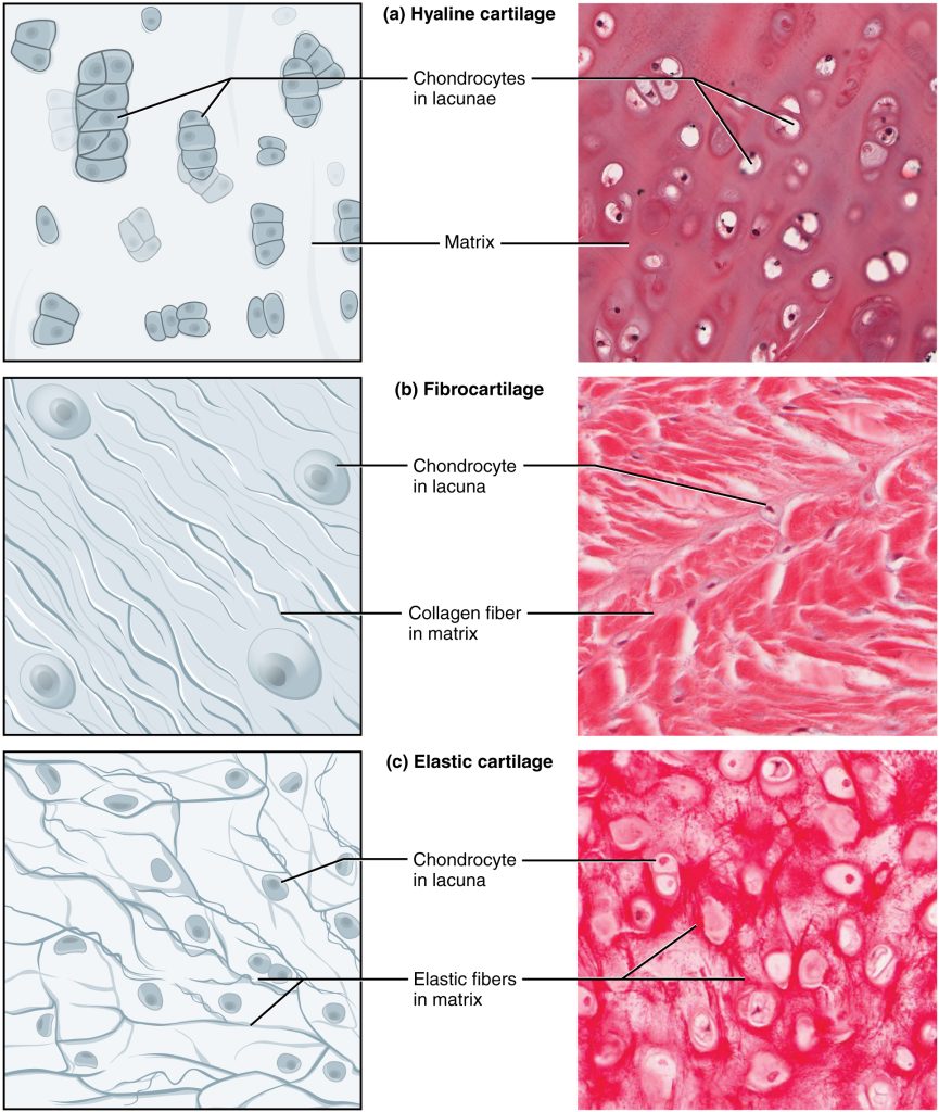
Cartilage has a relatively rigid matrix composed of protein fibers and proteoglycan aggregates. It contains mainly chondrocytes, within the lacunae. There are three types:
Hyaline cartilage with homogeneously distributed collagen fibers in the matrix and Chondrocytes in the lacunae
Fibrocartilage with homogeneously distributed collagen fibers but arranged in thick bundles and chondrocytes in the lacunae.
Elastic cartilage is similar to hyaline cartilage, but besides containing collagen fibers it also has elastin fibers.
-
Bone
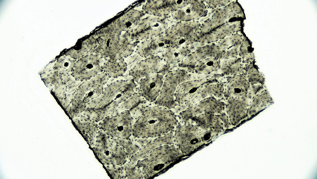
The bone cells, or osteocytes, are located in lacunae and surrounded by a mineralized matrix (hydroxyapatite). There are two types of bone; Spongy bone has spaces between bony trabeculae filled with bone marrow, and compact bone, which is more solid and composed of functional units called osteons.
-
Fluid Connective Tissue
Blood
The blood has a fluid matrix and specialized cells or formed elements of the blood; red blood cells, white blood cells and platelets.
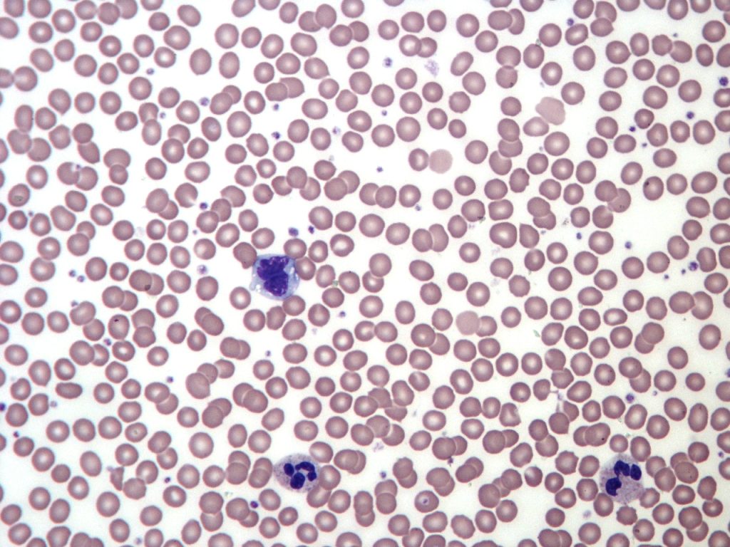
Creative Commons Attribution-Share Alike 3.0 Unported license.
Muscular Tissue
Muscle tissue has the ability to contract. There are three type of muscle tissue; Skeletal, smooth and cardiac muscle.
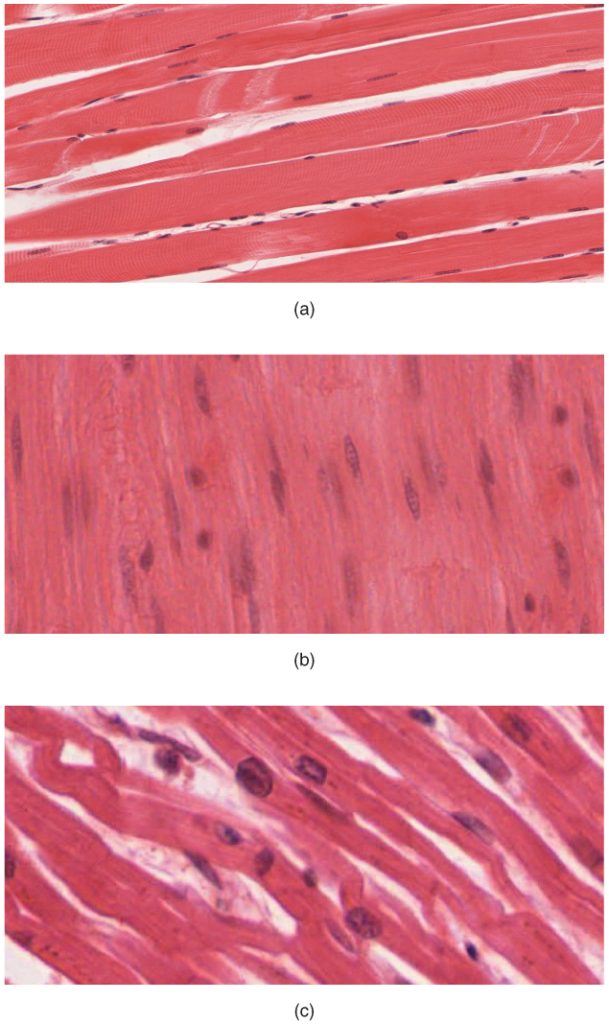
The body contains three types of muscle (x.s., 400x)
- skeletal muscle
- smooth muscle
- cardiac muscle
Virtual Microscope – use the QR code shown below to access the Virtual Microscope from University of Michigan. Use the +/- signs below the image to increase/decrease magnification. You may also scan the image – look at the top left of the main view, you will see a smaller window with a blue square, drag the square to change the area where you like to see. You can also move to a different area in the main view by dragging the small white hand shown there.

Skeletal Muscle
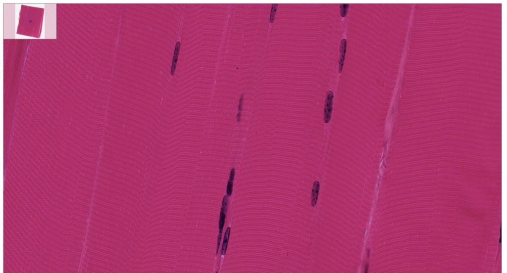
Skeletal muscle attaches to skeleton and is responsible for body movement. Skeletal muscle cells are long and cylindrically formed from fusion of many cells during development, therefore they contain many nuclei located at the periphery. This type of muscle has a banding pattern (striated) and is under voluntary control.
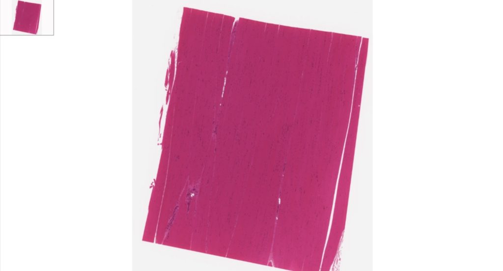
Then focus on the skeletal muscle fibers, long cylindrical, banded, multinucleated, nucleus located at the periphery.
Cardiac Muscle
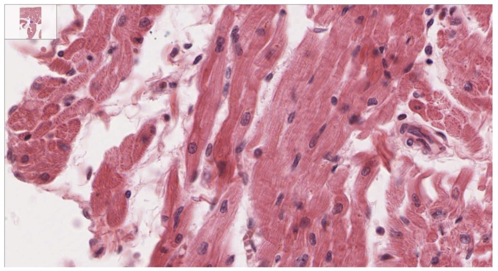
Cardiac muscle cells are short, branching cells with a single, central nucleus and gap junctions called intercalated disks. Cardiac muscle is only found in the heart and is responsible for pumping blood through the circulatory system. This type of muscle has a banding pattern (striated) and is under involuntary control.
Slide: 057_HISTO_40X
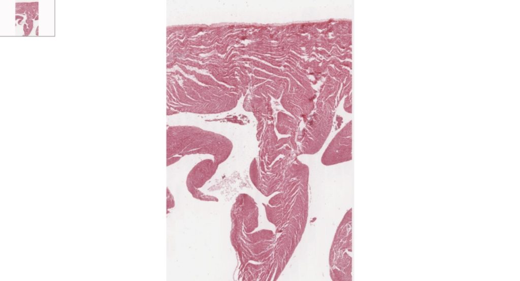
Then focus on cardiac cells
Smooth muscle
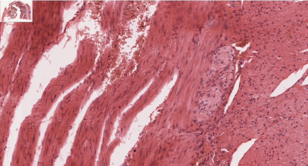
Smooth muscle is found in the walls of hollow organs, the iris of the eye, and other structures. The cells are tapered or spindle-shaped with a single, central nucleus. This type of muscle does not have a banding pattern (non-striated) and is under involuntary control.
Slide: 169_HISTO_40X
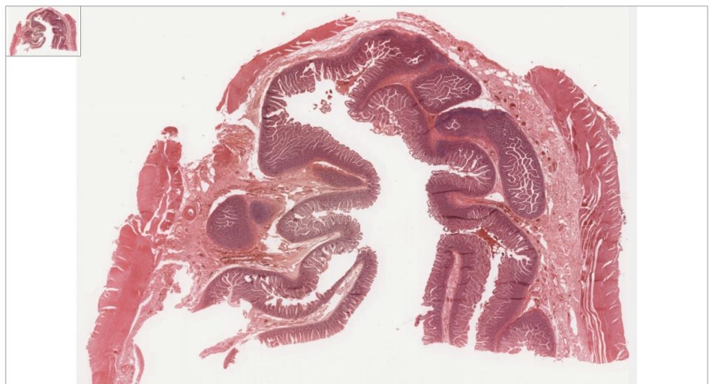
Focus on the smooth muscle, note tapered cells with central nucleus, no banding pattern.
Nervous Tissue
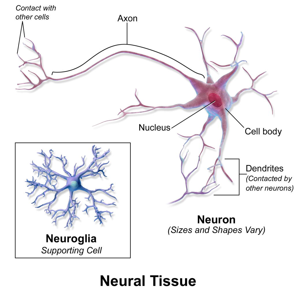
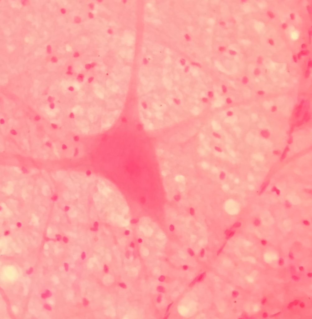
The nervous tissue is composed of two types of cells, neurons (functional units, capable of transmitting an action potential) and neuroglia that support, nourish and monitor the neurons, they are found in an average ratio of 1: 9 (neuron: neuroglia). These cells are contained in the organs of the nervous system. The Central Nervous System (CNS) is composed of the brain and the spinal cord, the Peripheral Nervous System (PNS) is composed of all tissue located outside of the CNS, cranial and peripheral nerves.
Neurons have distinct type of cell processes; dendrites and axons. Dendrites are located on the body of the neuron, they receive electrical impulses, and axons conduct them.
Virtual Microscope – use the QR code shown below to access the Virtual Microscope from University of Michigan. Use the +/- signs below the image to increase/decrease magnification. You may also scan the image – look at the top left of the main view, you will see a smaller window with a blue square, drag the square to change the area where you like to see. You can also move to a different area in the main view by dragging the small white hand shown there.

Neurons
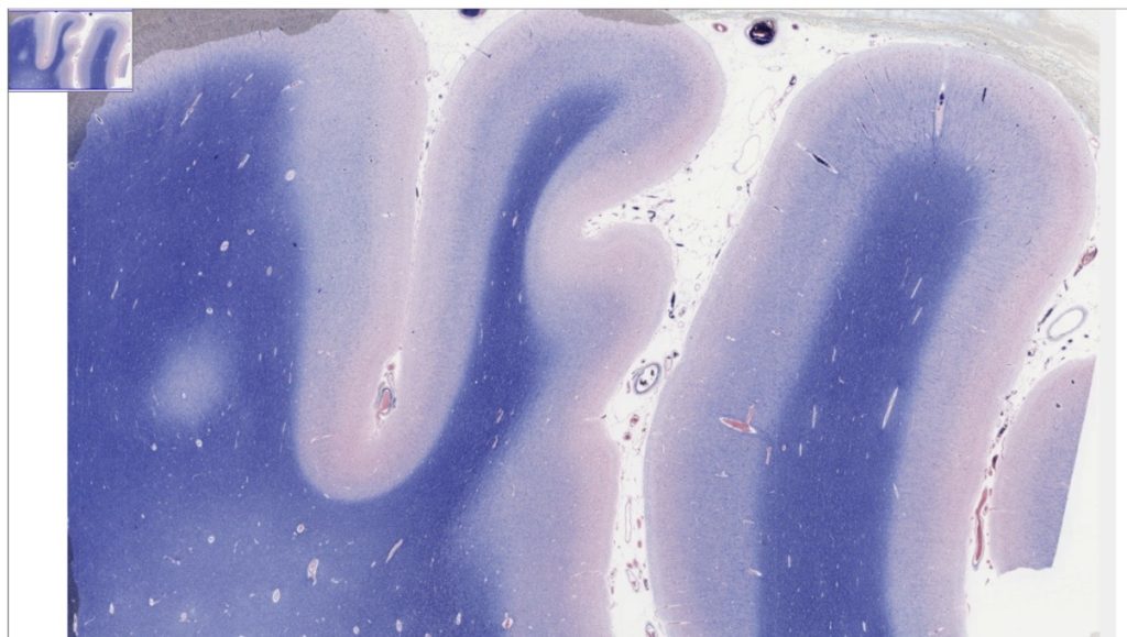
Cerebrum, axons and neuron cell bodies stained blue, 40X (white matter [stained dark blue], cerebral cortex, pyramidal cells, pyramid shaped neuron cell bodies of various size, dendritic tree not visible, axon goes to white matter], glial cells, sulcus [white line from top to bottom of this section] between gyri.
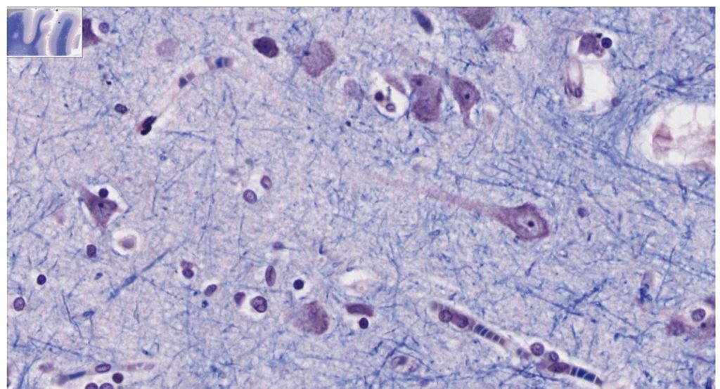
Then focus on an area where you can find neurons
Laboratory Activities
Epithelial Tissue Activity – Draw and Identify
Use a compound light microscope to examine organ slides provided in the lab. Follow the rules of proper use and maintenance of a microscope explained to you at the beginning of the semester. You will first need to locate the epithelial tissue; remember the slides we use are sections of organs, they may contain other types of tissues as well. Remember, epithelium has a free apical side.
Observe the epithelial tissue slides under scanning power to locate the area where the epithelium is located, microscopes are parfocal, you will change magnification without having to focus again, just use the micro adjustment know to fine tune the focusing. You do not need 100X objective for this activity.
For 100% online courses, you will be using a virtual microscope. Instead of focusing using the coarse and fine adjustment knobs, you will use the +/- signs below the image to increase or decrease magnification.
Observe and draw the following epithelial tissues slides:
- Simple squamous epithelium – you may use lung, cheek cells, or kidney (bowman’s capsule) slides.
- Simple cuboidal epithelium – you may use kidney (tubules) or thyroid gland slides
- Simple columnar epithelium – you may use stomach, duodenum, jejunum, ileum, colon or kidney slides.
- Pseudostratified columnar ciliated epithelium with goblet cells, respiratory epithelium – use Trachea slides
- Stratified squamous epithelium (non-keratinized) – you may use esophagus, vagina, or rectum slides
- Stratified squamous epithelium (keratinized) – you may use thin or thick skin slides
- Stratified cuboidal – use thin skin slides, locate the sebaceous glands and/or sweat glands in dermal layer.
- Stratified columnar epithelium – rare. We do not have this slide to look at, instead look at an image in your textbook.
- Transitional epithelium – you may use urinary bladder or ureter slides.
Note: Label the organelles you can identify, i.e, nucleus, cytoplasm, cell membrane, also label the basement membrane, and the free or luminal surface of the epithelium.






Identify the type of epithelial tissue seen under the microscopes
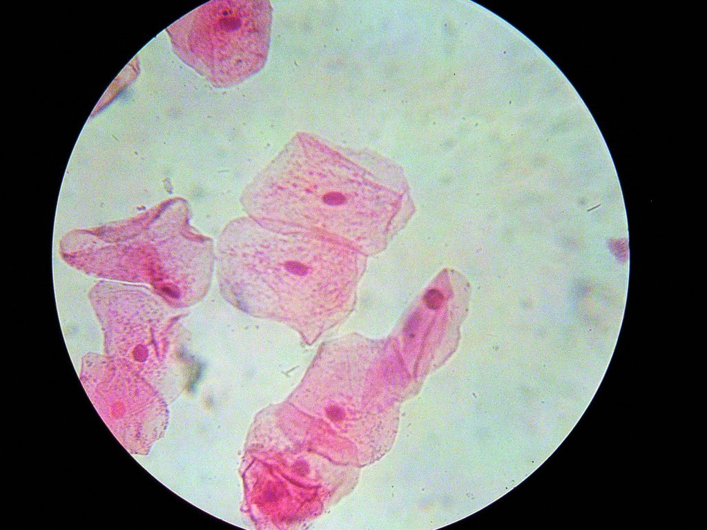
Tissue type:

Function:


Tissue type:

Function:

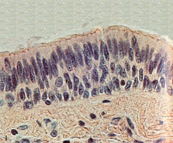
Tissue type:

Function:

Connective Tissue Activity – Draw and Identify
Use a compound light microscope to examine organ slides provided in the lab. Follow the rules of proper use and maintenance of a microscope explained to you at the beginning of the semester. You will first need to locate the connective tissue; remember the slides we use are sections of organs, they may contain other types of tissues as well.
Observe the connective tissue slides under scanning power to locate the area where the tissue microscopes are parfocal, you will change magnification without having to focus again, just use the micro adjustment know to fine tune the focusing. You do not need 100X objective for this activity.
Observe and draw the following connective tissues slides:
- Loose Areolar tissue, packing material. You need to find and label the fibroblast cells, ground substance, collagen and elastic fibers
- Adipose Tissue: Find cell membrane and nuclei.
- Reticular connective Tissue – found in lymph nodes and liver, find and label the reticular fibers and cells.
- Regular Dense CT- Collagenous tissue (or dense regular connective tissue), found in ligaments and tendons. You need to find and label collagen fibers and fibroblast cells
- Elastic connective tissue – (large arteries). You need to find and label circular layer of elastic fibers.
- Irregular Dense CT – found in dermis of skin. You need to find and label bundles of collagen fibers
- Hyaline cartilage – Find and label chondrocytes, lacunae, matrix
- Elastic cartilage – Find and label chondrocytes, lacunae, elastic fibers
- Fibrocartilage – Find and label chondrocytes, lacunae, collagen fibers
- Compact bone: Find and label – Osteons: lacunae, canaliculi, osteocyte, concentric lamellas
- Human blood smear. Find and label the formed elements of blood; RBC, red blood corpuscles (erythrocytes), WBC, white blood cells (leukocytes), neutrophils, eosinophils, basophils, monocytes and lymphocytes. Platelets (Thrombocytes). As you observe a blood smear under the microscope, draw all formed elements of the blood. Note the absence or presence of the nucleus, the nuclear shape, amount of cytoplasm, and presence or absence of granules in the cytoplasm.
Note: You will need to use immersion oil and 100X objective to see details of formed elements of blood.











Answer the following questions
- What are the 3 common characteristics of all connective tissue?

- Where in the body is each type of connective tissue found? What is their function? Make a table.
| Connective tissue type | Location |
- Name the formed elements of blood, state their function.

- Why does the RBCs lose their nucleus?

- What are the differences and similarities in WCBs types?

Muscle Tissue Activity – Draw and Identify
There are three types of muscle tissue, skeletal muscle, smooth muscle and cardiac muscle. You will learn how to identify each type of muscle tissue. You will first need to locate the muscle tissue; remember the slides we use are sections of organs, they may contain other types of tissues as well. Observe the muscle tissue slides under scanning power to locate the area where the tissue microscopes are parfocal, you will change magnification without having to focus again, just use the micro adjustment know to fine tune the focusing. You do not need 100X objective for this activity.
Observe and draw the following muscle tissues slides:
- Skeletal muscle (teased)– Find and label the striations, nuclei and cylindrical muscle cells (fibers)
- Smooth muscle (teased)– Find and label the nuclei and spindle shaped cells
- Cardiac muscle (intercalated disk stain) – Find and label the intercalated disks, striations, nuclei and branched cells.

Tissue Type: , Magnification __________X 
Tissue Type: , Magnification __________X 
Tissue Type: , Magnification __________X
Identify the type of muscle tissue seen under the microscopes
Demo Slide Activity
There are two microscopes set up at the instructor’s bench, identify each of them. Be specific. What is the function of each type of epithelial tissue you identified?
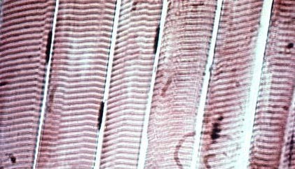
Microscope 1 – Tissue type:

Microscope 1 – Function:

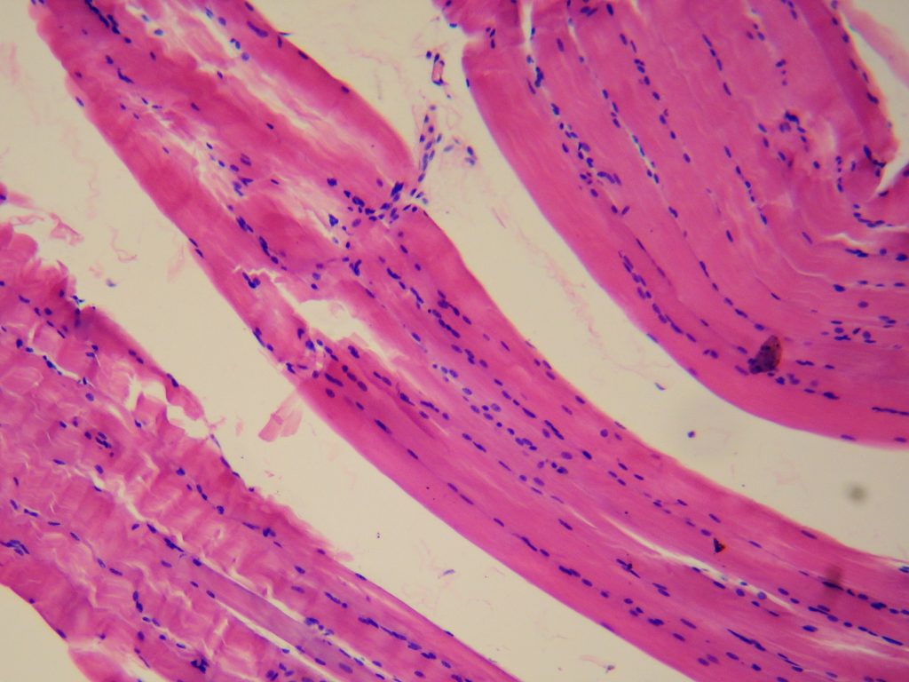
Microscope 2 – Tissue type:

Microscope 2 – Function:

Nervous Tissue Activity – Draw and Identify
- Microscopy
Use a compound light microscope to examine organ slides provided in the lab. You will first need to locate the nervous tissue; remember the slides we use are sections of organs, they may contain other types of tissues as well. Observe the nervous tissue slides under scanning power to locate the area where the tissue microscopes are parfocal, you will change magnification without having to focus again, just use the micro adjustment know to fine tune the focusing. You do not need 100X objective for this activity. Observe and draw a multipolar neuron smear slide or motor neuron, spinal cord smear, which ever one is available
Find and label the following structures:
- Motor neurons, cell body, nucleus, Nissl bodies, dendrites and axons.
- Neuroglia cell bodies

Tissue Type: , Magnification __________X
Identification of structures using the 3D neuron anatomical model. identify the following:
- Cell body
- Nucleus
- Nissl bodies
- Dendrites
- Axons
- Myelin sheath
- Node of Ranvier
- Internodes
- Synaptic terminals – Telodendria
Label the components of a typical motor neuron
Review
Tissue type
| Epithelium: | Comments | Location |
| Epithelium, simple squamous | ||
| Epithelium, stratified squamous | ||
| Epithelium, simple cuboidal | ||
| Epithelium, pseudostratified ciliated columnar | ||
| Epithelium, simple columnar | ||
| Transitional epithelium |
| Loose connective Tissue: | Comments | Location |
| Areolar tissue | ||
| Adipose tissue | ||
| Reticular tissuel |
| Dense connective Tissue: | Comments | Location |
| Dense regular | ||
| Dense irregular |
| Supportive connective Tissue: Cartilage | Comments | Location |
| Fibrocartilage | ||
| Elastic cartilage (yellow cartilage) | ||
| Hyaline cartilage |
| Supportive connective Tissue: Bone | Comments | Location |
| Compact | ||
| Spongy bone |
| Fluid connective Tissue: | Comments | Location |
| Blood |
| Muscular Tissue: | Comments | Location |
| Skeletal muscle | ||
| Cardiac muscle, intercalated disks | ||
| Smooth muscle |
| Nervous Tissue: | Comments | Location |
| Neuronal smear (neurons and neuroglia) |
Media Attributions
- Compact bone © Elchiaru, CC BY 4.0 , via Wikimedia Commons is licensed under a CC BY (Attribution) license
- Types of epithelium © Wikipedia is licensed under a CC BY (Attribution) license
- Table Summary of Epithelial © Wikipedia is licensed under a CC BY (Attribution) license
- Simple squamous epithelium © Maria Carles adapted by Wikipedia is licensed under a CC BY-SA (Attribution ShareAlike) license
- QR code V Microscope © Maria Carles is licensed under a CC BY-SA (Attribution ShareAlike) license
- Simple cuboidal epithelium © Michigan Medical School adapted by Maria Carles is licensed under a CC BY-SA (Attribution ShareAlike) license
- Simple squamous cell © Michigan Medical School adapted by Maria Carles is licensed under a CC BY-NC-SA (Attribution NonCommercial ShareAlike) license
- Intradermal Nevus © Wikipedia is licensed under a CC BY-SA (Attribution ShareAlike) license
- Thin skin 40X © Michigan Medical School adapted by Maria Carles is licensed under a CC BY-SA (Attribution ShareAlike) license
- Epidermal layer © Maria Carles adapted by Michigan Medical School is licensed under a CC BY-NC-SA (Attribution NonCommercial ShareAlike) license
- Thyroid histology © Wikipedia is licensed under a CC BY (Attribution) license
- Kidney, human © Michigan Medical School adapted by Maria Carles is licensed under a CC BY-SA (Attribution ShareAlike) license
- Cuboidal section of tubules © Maria Carles adapted by Michigan Medical School is licensed under a CC BY-NC-SA (Attribution NonCommercial ShareAlike) license
- Gastric heterotopia in the duodenum © Wikipedia is licensed under a CC BY (Attribution) license
- Colon © Michigan Medical School is licensed under a CC BY-SA (Attribution ShareAlike) license
- Colon, H&E, 40X © Michigan Medical School adapted by Maria Carles is licensed under a CC BY-SA (Attribution ShareAlike) license
- Pseudostratified columnar ciliated epithelium © Michigan Medical School adapted by Maria Carles is licensed under a CC BY-SA (Attribution ShareAlike) license
- Trachea H&E © Michigan Medical School adapted by Maria Carles is licensed under a CC BY-SA (Attribution ShareAlike) license
- Trachea H&E, 40X © Michigan Medical School adapted by Maria Carles is licensed under a CC BY-SA (Attribution ShareAlike) license
- Epithelial tissues transitional © Wikipedia is licensed under a CC BY (Attribution) license
- Non-distended bladder © Michigan Medical School adapted by Maria Carles is licensed under a CC BY-SA (Attribution ShareAlike) license
- Transitional epithelium © Maria Carles adapted by Michigan Medical School is licensed under a CC BY-NC-SA (Attribution NonCommercial ShareAlike) license
- Connective tissues © Wikipedia is licensed under a CC BY (Attribution) license
- Tejidos conjuntivos © Wikipedia is licensed under a CC BY (Attribution) license
- Connective Tissue Loose Aerolar © Wikipedia is licensed under a CC BY (Attribution) license
- Yellow adipose tissue © Wikipedia is licensed under a CC BY (Attribution) license
- Connective tissue reticular © Wikipedia is licensed under a CC BY (Attribution) license
- Dense connective tissue © Wikipedia is licensed under a CC BY (Attribution) license
- Collagen fibers © Maria Carles adapted by Wikipedia is licensed under a CC BY-SA (Attribution ShareAlike) license
- Human elastic tissue © Wikipedia is licensed under a CC BY (Attribution) license
- Types of cartilage © Wikipedia is licensed under a CC BY (Attribution) license
- Tissue compact bone © Wikipedia is licensed under a CC BY (Attribution) license
- Adult blood smear © Wikipedia is licensed under a CC BY (Attribution) license
- Skeletal smooth cardiac © wikipedia is licensed under a CC BY (Attribution) license
- Skeletal Muscle © Michigan Medical School is licensed under a CC BY-SA (Attribution ShareAlike) license
- Skeletal muscle fibers © Maria Carles adapted by Michigan Medical School is licensed under a CC BY-NC-SA (Attribution NonCommercial ShareAlike) license
- Cardiac Muscle © Michigan Medical School adapted by Maria Carles is licensed under a CC BY-SA (Attribution ShareAlike) license
- Cardiac muscle © Maria Carles adapted by Michigan Medical School is licensed under a CC BY-NC-SA (Attribution NonCommercial ShareAlike) license
- Smooth muscle © Michigan Medical School adapted by Maria Carles is licensed under a CC BY-NC-SA (Attribution NonCommercial ShareAlike) license
- Smooth muscle, 400X
- Neural Tissue © Wikipedia is licensed under a CC BY (Attribution) license
- Neuron section © Wikipedia is licensed under a CC BY (Attribution) license
- Neuron cells © Michigan Medical School adapted by Maria Carles is licensed under a CC BY-NC-SA (Attribution NonCommercial ShareAlike) license
- Neurons © Michigan Medical School adapted by Maria Carles is licensed under a CC BY-NC (Attribution NonCommercial) license
- Blank circle
- Blank circle
- Cheek cells stained © Wikipedia is licensed under a CC BY-SA (Attribution ShareAlike) license
- Columnar Estratificado © Wikipedia is licensed under a CC BY-SA (Attribution ShareAlike) license
- Blank circle
- Blank circle
- Blank circle
- Blank circle
- Blank circle
- Blank circle
- Blank circle
- Blank circle
- Blank circle
- Tissue type © Wikipedia is licensed under a CC BY-SA (Attribution ShareAlike) license
- Tissue type 2 © Wikipedia is licensed under a CC BY-SA (Attribution ShareAlike) license
- Neuron hand tuned © Wikipedia is licensed under a CC BY-SA (Attribution ShareAlike) license

