The Integumentary System Anatomy
The Integumentary System
Objectives:
- Identify components of the integumentary system
- List the two types of skin
- Describe macro and microscopic anatomy of both types of skin
- List accessories structures of the skin
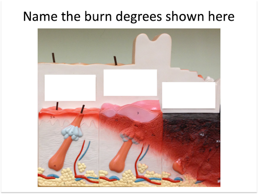
- Associate the layers of skin and hypodermis with burn degrees
Introduction
The integumentary system is the largest organ of the human body. It is composed of four types of tissue, epithelial, connective, muscular and nervous tissues. The structures of the integumentary system include the skin, and its accessory organs; hairs, nails, accessory glands, sensory receptors. The integumentary system protects the body from dehydration, absorption of nutrients, excretion of wastes, and regulation of body temperature. It is also the attachment site of certain muscles and contains sensory receptors to detect pain, sensation, pressure, and temperature and it is also involved in synthesis of vitamin D.
In this lab you will learn to identify the four types of tissues that compose the integumentary system and correlate them with their function.
Part 1 – Anatomy of the Skin
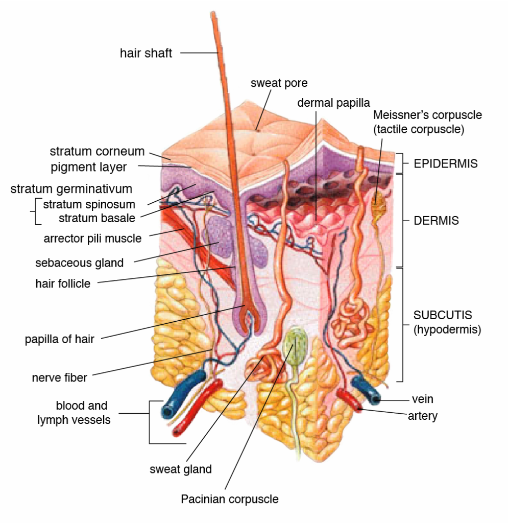
The skin is also called the cutaneous membrane. There are two types of skin, thin skin that is covered with hair (also contains sebaceous glands) and thick skin that has no hair.
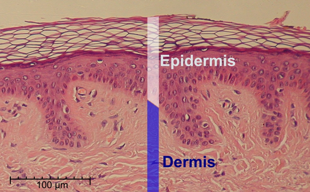
The skin, regardless of the type is composed of two layers; the epidermis and the dermis. It has an associated layer below the dermis, called the hypodermis.
Epidermis
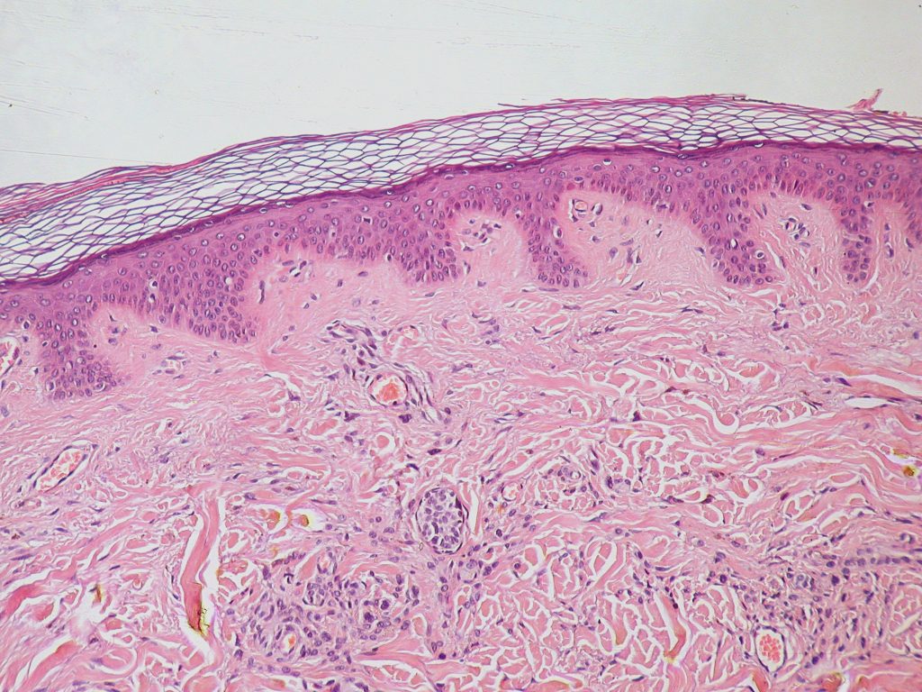
The epidermis is the top layer of skin. It is avascular, meaning, it does not contain blood vessels. It consists of layers of stratified squamous epithelium, and contains four types of cells (keratinocytes, melanocytes, Merkel cells, and Langerhans cells). The keratinocytes are the predominant cell type in the epidermis, they produce keratin, a fibrous protein that protects skin from mechanical stress. The majority of the skin on the body is keratinized. The only skin on the body that is non-keratinized is the lining of mucous membranes, such as the inside of the mouth.
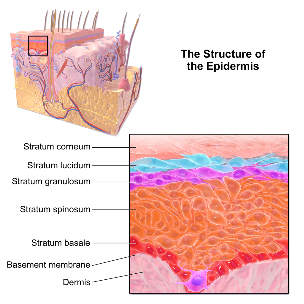
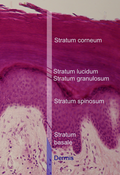
The epidermis contains 4-5 layers or strata of stratified squamous epithelium.
We will describe them from deep to superficial.
- Stratum basale: Deepest layer of the epidermis, it is composed mainly of one layer of proliferating and non-proliferating keratinocytes, attached to the basement membrane by hemidesmosomes. Melanocytes are present in this layer, connected to numerous keratinocytes through dendrites. Merkel cells are also found in the stratum basale (large numbers of touch-sensitive receptors are found in fingertips and lips).
- Stratum spinosum: Keratinocytes become connected through desmosomes and start to produce lamellar bodies, from within the Golgi, enriched in polar lipids, glycosphingolipids, free sterols, phospholipids and catabolic enzymes. Langerhans cells (immunologically active cells) and melanin are found in the middle of this layer.
- Stratum granulosum: layer in which keratinocytes lose their nuclei and their cytoplasm appears granular. Lipids, contained into those keratinocytes within lamellar bodies, are released into the extracellular space through exocytosis to form a lipid barrier.
- Stratum lucidum: A clear/translucent layer of dead keratinocytes, only present in thick skin.
- Stratum corneum: Superficial layer of cells, composed of 10 – 30 layers anucleated keratinocyte, with the palms and soles having the most layers. Most of the barrier functions of the epidermis localize to this layer.
Dermis
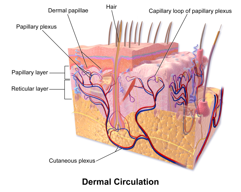
The dermis is the middle layer of skin, composed of areolar connective tissue and dense irregular connective tissue. The dermis has two layers. The papillary layer (superficial) which consists of the areolar connective tissue. The reticular layer (deep) which consists of the dense irregular connective tissue. These layers serve to give elasticity and flexibility to the integument. The dermal layer provides a site for the blood vessels, free nerve endings, pressure receptors (Pacinian corpuscles), sweat glands, sebaceous glands, hair follicles, arrector pili muscles.
Hypodermis
The hypodermis, also known as the subcutaneous layer, is a layer beneath the skin. It invaginates into the dermis and is attached to the latter, immediately above it, by collagen and elastin fibers. It is essentially composed of a type of cell known as adipocytes specialized in accumulating and storing fats. These cells are grouped together in lobules separated by connective tissue. The hypodermis acts as an energy reserve. The fat contained in the adipocytes can be brought into circulation, via the venous route, during intense effort or when there is a lack of energy providing a source for energy production. The hypodermis participates passively in thermoregulation because the fat stored in adipocytes acts as a heat insulator.
Part 2 – Anatomy of the accessory organs of the skin
Hair
Hair is a protein filament that grows from follicles found in the dermis. The human body, apart from areas of glabrous skin, is covered in follicles which produce thick terminal and fine vellus hair.
Hair fibers have a structure consisting of several layers, starting from the outside:
- Cuticle, outer covering, which consists of several layers of flat, thin cells laid out overlapping one another as roof shingles
- Cortex, which contains the keratin bundles in cell structures that remain roughly rod-like. The highly structured and organized cortex, or second of three layers of the hair, is the primary source of mechanical strength and water uptake. The cortex contains melanin, which colors the fiber based on the number, distribution and types of melanin granules. The shape of the follicle determines the shape of the cortex, and the shape of the fiber is related to how straight or curly the hair is. People with straight hair have round hair fibers. Oval and other shaped fibers are generally more wavy or curly.
- Medulla, a disorganized and open area at the fiber’s center. The innermost region of the hair.
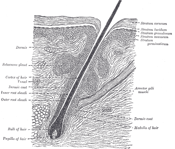
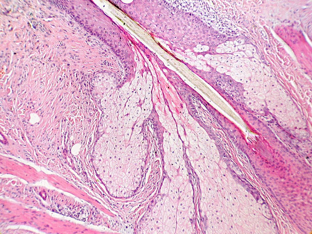
Hair growth begins inside the hair follicle. The only “living” portion of the hair is found in the follicle. The hair that is visible is the hair shaft, which exhibits no biochemical activity and is considered “dead”. The base of a hair’s root (the “bulb”) contains the cells that produce the hair shaft. Other structures of the hair follicle include the oil producing sebaceous gland which lubricates the hair and the arrector pili muscles, which are responsible for causing hairs to stand up. In humans with little body hair, the effect results in goose bumps.
Section of skin, showing the epidermis and dermis; a hair in its follicle; the Arrector pili muscle; sebaceous glands.
Nails
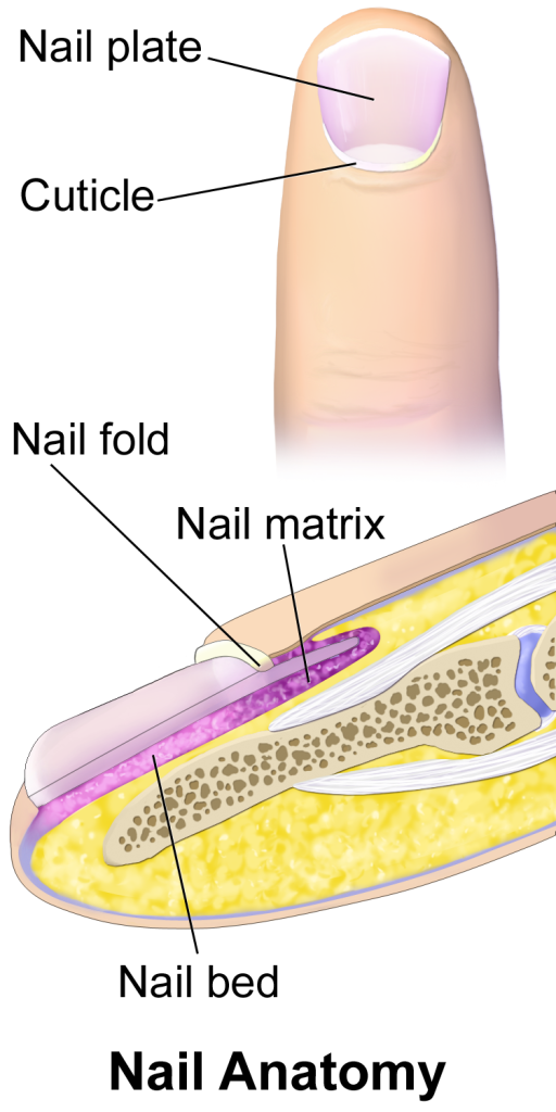
Nails are composed like hair mostly of dead keratinocytes, the protein keratin stiffens epidermal tissue to form fingernails. Nails grow from a thin area called the nail matrix at an average of 1 mm per week.
A nail is composed of a flat nail plate surrounded by nail folds. The proximal nail fold grows over the bail plate forms a structure called the cuticle or eponychium. Near the eponychium you can find a thickened area of the nail plate that looks like a half a moon (crescent-shape), this area is called the lunula.
Laboratory Activity – Skin
Macroscopic Anatomy of Skin
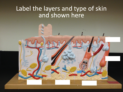
Use the Anatomical Model of Skin (Denoger Geppert #185) to identify parts of the skin (thick and thin skin):
- Epidermis (with all layers), Dermis and the associated layer the hypodermis and types of skin (thin or thick). Highlighted in yellow needs to be added.
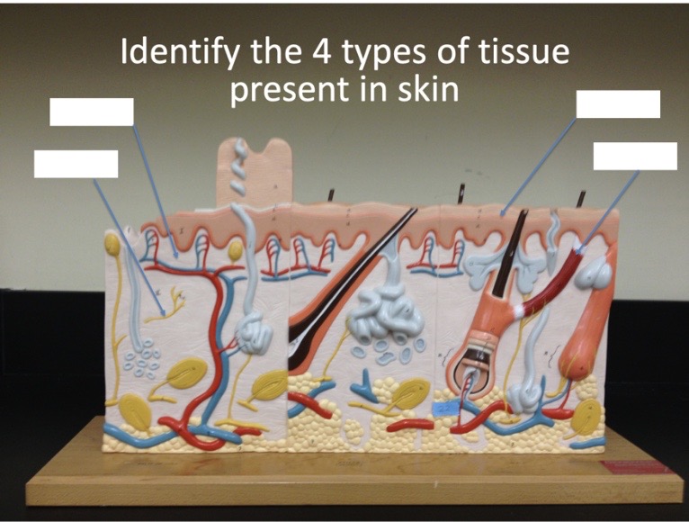
- Identify 4 types of tissue: Epithelial, muscular, connective and nervous using this model.
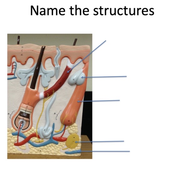
- Identify sweat glands, sebaceous glands, blood vessels, free nerve endings, Meisner corpuscles, arrector pili muscle, hair follicle, hair plexus.
- Describe burn degrees and associate them with layers of the skin and hypodermic layer.
First degree:
Second degree:
Third degree:
Microscopy Anatomy of Skin
Observe microscope slides of different types of skin:
- Human Thick Skin (Pigmented).
Draw what you see and label the following structures:
- Epidermis (all layers)
- stratum corneum
- stratum granulosum
- stratum spinosum
- stratum basale (note the pigmentation in this layer). What is the type of cells that produce the pigmentation seen here?
- Dermis
- Subcutaneous layer
Note: for 100% online courses use UM Virtual Microscope.

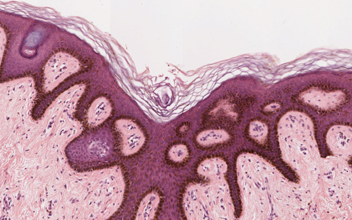
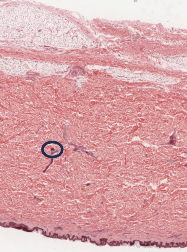
Now look at slide 105-2, Pigmented skin at lowest magnification and focus on the area of the dermis delineated with a circle, what type of tissue do you see? What do you think this is?
- White human skin.
Draw and label the following:
-
- Epidermis (all layers)
- stratum corneum
- stratum granulosum
- stratum spinosum
- stratum basale
- Dermis
- Subcutaneous layer
- Epidermis (all layers)
Note: for 100% online courses use UM Virtual Microscope.
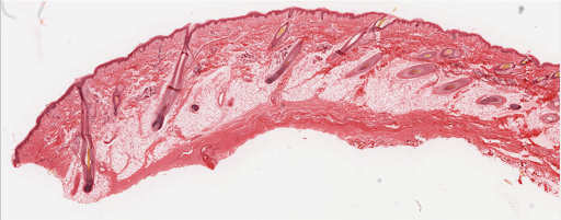
Now Zoom in to high magnification and moving around locate draw and label the following structures:
- hair follicles
- external root sheath
- Sebaceous glands
- Arrector pili
- Sweat gland ducts
- Adipose tissue
- Thick Skin.
Draw and label the following:
-
- Epidermis
- stratum corneum
- stratum lucidum
- stratum granulosum
- stratum spinosum
- stratum basale
- Dermis
- Ducts of sweat glands
- Subcutaneous layer
- Epidermis
Note: for 100% online courses use UM Virtual Microscope.

slide # 112N – Thick skin, sole of foot
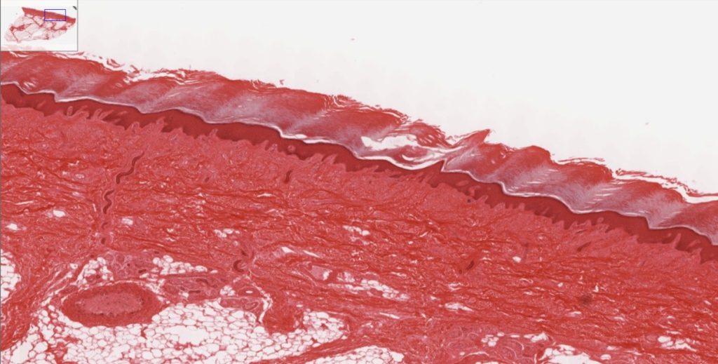
Note: to be able to see the layers of the epidermis, you need to Zoom in.
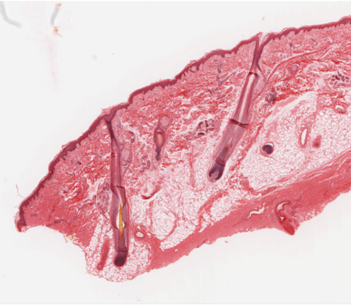
- Human Scalp c.s – Examine a longitudinal section of a hair follicle
Draw and label the following; hair shaft, hair follicle, bulb. dermal papilla, sebaceous gland, sweat gland and the region of cell division. Go to UM virtual microscope – Skin and Mammary glands section, Slide#107, Scalp, hair, H&E
-
- Download the image for labeling.
-
-
- Using low power, locate and label the following structures:
- Hair follicle
- Sebaceous glands
-
-
- Using high power locate the following structures:
-
-
- External root sheath of the hair follicle
- Root of hair follicle
- Hair shaft
- Arrector pili muscle
-
- Anatomy of a nail
- Go to UM Virtual microscope slide # 108 20X– Fetal finger
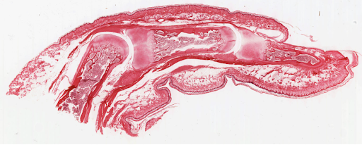
- Label the following structures:
- Go to UM Virtual microscope slide # 108 20X– Fetal finger
-
-
- Nail fold
- Nail matrix
- NAIL bed
- Distal medial and proximal phalanges
-
-
- Zoom in and locate the following tissues:
-
-
- hyaline cartilage
- bone
- epidermis
- dermis
-
Apply what you have learned
Answer the following questions. You may need to use the textbook as reference to answer these questions.
- What is the predominant tissue type in the epidermis?

- What is the predominant tissue type in the dermis?

- What is the predominant tissue type in the hypodermis?

- Were you able to identify the melanocytes? In which layer do you see them? What is their function?

- Which other structures can you identify within the dermis?

- What type of tissue makes the ducts of the sweat glands? Did you find one?

- What type of tissue is present in the dermis of the skin?

- List the layers of a hair, as seen from a cross section of hair.

Media Attributions
- Skin © Wikipedia is licensed under a CC BY-SA (Attribution ShareAlike) license
- Human Skin © Wikipedia is licensed under a CC BY (Attribution) license
- Epidermis delimited © Wikipedia is licensed under a Public Domain license
- Epidermis structure © Wikipedia is licensed under a CC BY-SA (Attribution ShareAlike) license
- Epidermal layers © Wikipedia is licensed under a CC BY-SA (Attribution ShareAlike) license
- Dermal circulation © Wikipedia is licensed under a CC BY-SA (Attribution ShareAlike) license
- Diagram from Gray’s Anatomy © Wikipedia is licensed under a CC BY-SA (Attribution ShareAlike) license
- Sebaceous glands into hair shaft © Wikipedia is licensed under a Public Domain license
- Finger nail anatomy © Wikipedia is licensed under a CC BY-SA (Attribution ShareAlike) license
- Label the layers © Maria Carles is licensed under a CC BY-SA (Attribution ShareAlike) license
- ID 4 types sof tissue © Maria Carles is licensed under a CC BY-SA (Attribution ShareAlike) license
- Name the structures © Maria Carles is licensed under a CC BY-SA (Attribution ShareAlike) license
- Name the burn degrees © Maria Carles is licensed under a CC BY-SA (Attribution ShareAlike) license
- QR code V Microscope © Maria Carles is licensed under a CC BY-SA (Attribution ShareAlike) license
- Thin or thick skin © Maria Carles adapted by Michigan Medical School is licensed under a CC BY-NC-SA (Attribution NonCommercial ShareAlike) license
- White human skin © Maria Carles adapted by Michigan Medical School is licensed under a CC BY-NC-SA (Attribution NonCommercial ShareAlike) license
- Thin skin © Maria Carles adapted by Michigan Medical School is licensed under a CC BY-NC-SA (Attribution NonCommercial ShareAlike) license
- Slide 112 © Maria Carles adapted by Michigan Medical School is licensed under a CC BY-NC-SA (Attribution NonCommercial ShareAlike) license
- Human scalp © Maria Carles adapted by Michigan Medical School is licensed under a CC BY-NC-SA (Attribution NonCommercial ShareAlike) license
- Anatomy of a nail © Maria Carles adapted by Michigan Medical School is licensed under a CC BY-NC-SA (Attribution NonCommercial ShareAlike) license

