The Muscular Tissue
Muscle Tissue Laboratory Activity
Objectives
- Describe the three types of muscle tissue and describe the basic function of each.
- Describe the histological appearance of the 3-muscle tissue types; skeletal, smooth and cardiac muscle.
- Identify each type of muscle tissue in microscope preparations and identify the microanatomy of the skeletal muscle fibers.
Introduction:
Muscle tissue is composed of cells that have the ability to shorten/contract in order to produce movement. The muscle cells, also called muscle fibers are long and slender. They are arranged in bundles or layers that are surrounded by connective tissue. Actin and myosin are the main contractile proteins in muscular tissue. Muscle tissue formed during embryonic development through a process known as myogenesis.
Muscle tissue is categorized into skeletal muscle tissue (attached to the skeleton), smooth muscle tissue (found inside of hollow organs and blood vessels, and cardiac muscle tissue (only found in the heart).
Skeletal muscle fibers are long, cylindrical, multinucleated (due to fusion of many cells during development), showing a banding pattern or striations, and are under voluntary control.
Smooth muscle cells are shorter, tapered or spindle shaped, have a central single, and lack striations. They are under involuntary control.
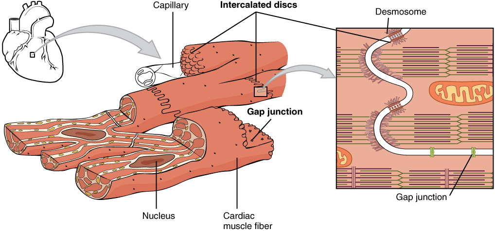
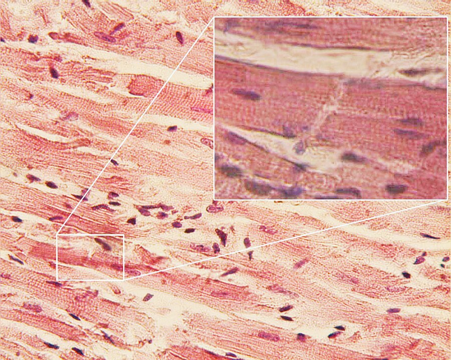
Cardiac muscles are short branched, one-two nucleus per cell, with striations, and modified gap junctions called intercalated disks. They are under involuntary control.
These muscle types are activated both through interaction of the central nervous system and endocrine (hormonal) activation. Skeletal muscle only contracts voluntarily, upon influence of the central nervous system. Reflexes are a form of nonconscious activation of skeletal muscles, but nonetheless arise through activation of the central nervous system, albeit not engaging cortical structures until after the contraction has occurred.
A schematic diagram of the different types of muscle cells
1) Skeletal muscle cells are long tubular cells with striations (3) and multiple nuclei (4). The nuclei are embedded in the cell membrane (5) so that they are just inside the cell. This type of tissue occurs in the muscles that are attached to the skeleton. Skeletal muscles function in voluntary movements of the body.
2) Smooth muscle cells are spindle shaped (6), and each cell has a single nucleus (7). Unlike skeletal muscle, there are no striations. Smooth muscle acts involuntarily and functions in the movement of substances in the lumens. They are primarily found in blood vessel walls and walls along the digestive tract.
3) Cardiac muscle cells branch off from each other, rather than remaining along each other like the cells in the skeletal and smooth muscle tissues. Because of this, there are junctions between adjacent cells (9). The cells have striations (8), and each cell has a single nucleus (10). This type of tissue occurs in the wall of the heart and its primary function is for pumping blood
Smooth Muscle
Smooth muscle is an involuntary non-striated muscle. It is divided into two subgroups: the single-unit (unitary) and multiunit smooth muscle. Within single-unit cells, the whole bundle or sheet contracts as a syncytium (i.e. a multinucleate mass of cytoplasm that is not separated into cells). Multiunit smooth muscle tissues innervate individual cells; as such, they allow for fine control and gradual responses, much like motor unit recruitment in skeletal muscle.
Smooth muscle is found within the walls of blood vessels (such smooth muscle specifically being termed vascular smooth muscle) such as in the tunica media layer of large (aorta) and small arteries, arterioles and veins. Smooth muscle is also found in lymphatic vessels, the urinary bladder, uterus (termed uterine smooth muscle), male and female reproductive tracts, gastrointestinal tract, respiratory tract, arrector pili of skin, the ciliary muscle, and iris of the eye. The structure and function is basically the same in smooth muscle cells in different organs, but the inducing stimuli differ substantially, in order to perform individual effects in the body at individual times. In addition, the glomeruli of the kidneys contain smooth muscle-like cells called mesangial cells.

The dense bodies and intermediate filaments are networked through the sarcoplasm, which cause the muscle fiber to contract.
Cardiac Muscle
Cardiac muscle (also called heart muscle or myocardium) is one of three types of vertebrate muscles, with the other two being skeletal and smooth muscles. It is an involuntary, striated muscle that constitutes the main tissue of the walls of the heart. The myocardium forms a thick middle layer between the outer layer of the heart wall (the epicardium) and the inner layer (the endocardium), with blood supplied via the coronary circulation. It is composed of individual heart muscle cells (cardiomyocytes) joined together by intercalated discs, encased by collagen fibres and other substances that form the extracellular matrix.
Cardiac muscle contracts in a similar manner to skeletal muscle, although with some important differences. An electrical stimulation in the form of an action potential triggers the release of calcium from the cell’s internal calcium store, the sarcoplasmic reticulum. The rise in calcium causes the cell’s myofilaments to slide past each other in a process called excitation contraction coupling.
Muscle tissue type Activities
Virtual Microscopy (for 100% online courses)
Go to University of Michigan Virtual Microscope, search for muscles, then choose the following slides:
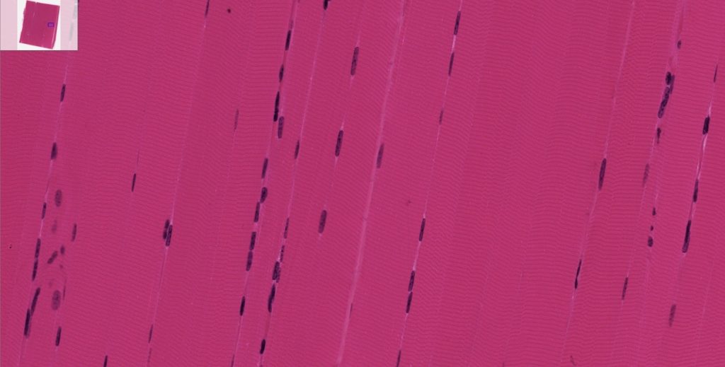
- Skeletal Muscle – Slide: #058 Thin section
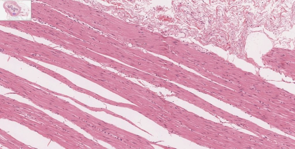
- Smooth Muscle – Slide: # 029-1
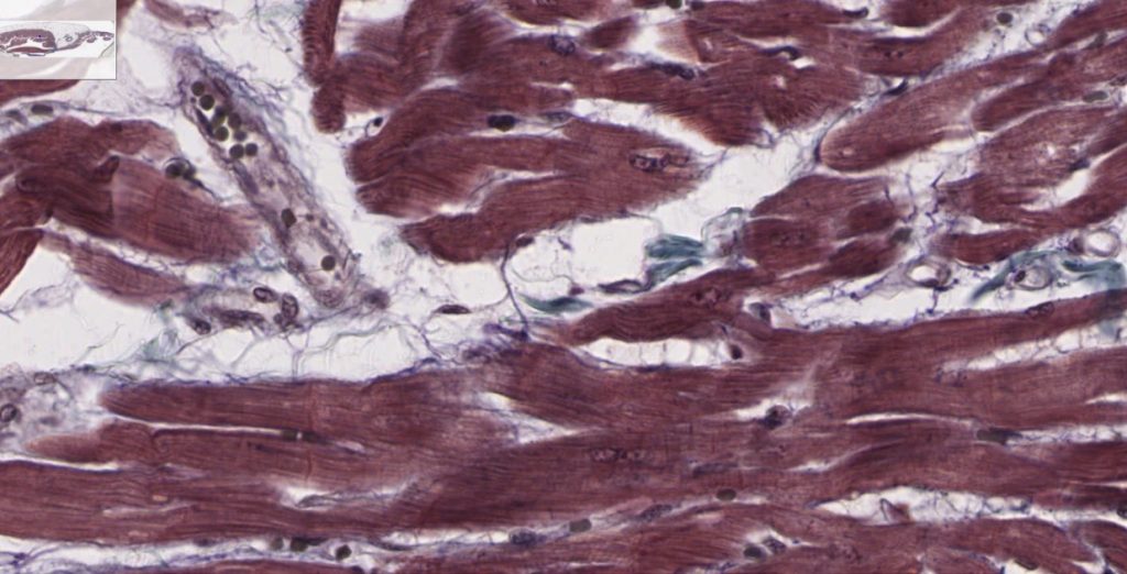
- Cardiac Muscle – Slide: #098-N
Instructions:
Once you have searched for the muscle’s slides, click on the slide number described above.
The first image you see is at scanning power level, you will need to increase magnification using the plus/minus signs until you see the area shown here.
Remember, these are slides of organs and contain more than one type of tissue, you need to locate the specific tissue, i.e. muscles (skeletal, smooth and cardiac). Locate the area where each of the muscle tissues (individual slides) are located draw and label them.
- Skeletal muscle – Slide: #058 Thin sectionDraw what you see in the space provided. Find and label the striations, peripheral nuclei and cylindrical muscle cells (fibers).
- Smooth muscle – Slide: # 029-1Draw what you see in the space provided. Find nuclei and spindle shaped cells.

- Cardiac muscle – Slide: #098-NDraw what you see in the space provided. Find intercalated disks, striations, nuclei and branched cells.

Light Microscopy (for hybrid and F-to-F courses)
Obtain muscle slides from your instructor. Look under the microscope at scanning (to locate the tissue), the change to low power and high power. Draw and label the structures of the following muscular tissues slides:
- Skeletal muscle (teased)Move around the microscope slide (move the mechanical stage once you have placed the slide in it and secure it with the clip) to locate the area where skeletal muscle is located (remember you are looking at organ slides, made of more than one type of tissue).
You should look for an area that looks like this:
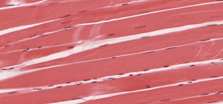
(a) skeletal muscle, LM × 1600. Draw what you see in the space provided. Find and label the striations, peripheral nuclei and cylindrical muscle cells (fibers).

- Smooth muscle (teased)Move around the microscope slide (move the mechanical stage once you have placed the slide in it and secure it with the clip) to locate the area where skeletal muscle is located (remember you are looking at organ slides, made of more than one type of tissue).
You should look for an area that looks like this:

Smooth muscle, LM × 1600. Draw what you see in the space provided. Find nuclei and spindle shaped cells.

- Cardiac muscle (intercalated disk stain)Move around the microscope slide (move the mechanical stage once you have placed the slide in it and secure it with the clip) to locate the area where skeletal muscle is located (remember you are looking at organ slides, made of more than one type of tissue).
You should look for an area that looks like this:
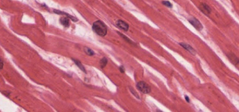
Cardiac muscle, LM × 1600. Draw what you see in the space provided. Find intercalated disks, striations, nuclei and branched cells.

The Sarcomere
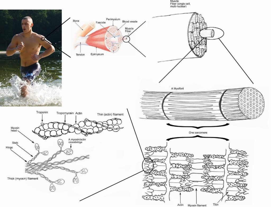
Use the anatomical Model of Skeletal muscle available in the lab.
Identify the following areas of the sarcomere:
I band, A band, H band, Z line, M line, thin and thick filaments and Zone of overlap.
Media Attributions
- Muscle Tissue © Wikipedia is licensed under a CC BY-SA (Attribution ShareAlike) license
- Smooth muscle contraction © By OpenStax - https://cnx.org/contents/FPtK1zmh@8.25:fEI3C8Ot@10/Preface, CC BY 4.0, https://commons.wikimedia.org/w/index.php?curid=30015054 is licensed under a CC BY-NC-SA (Attribution NonCommercial ShareAlike) license
- Cardiac muscle © By OpenStax - https://cnx.org/contents/FPtK1zmh@8.25:fEI3C8Ot@10/Preface, CC BY 4.0, https://commons.wikimedia.org/w/index.php?curid=30015048 is licensed under a CC BY-NC-SA (Attribution NonCommercial ShareAlike) license
- Cardiac muscle © By Dr. S. Girod, Anton Becker - Own work, CC BY 2.5, https://commons.wikimedia.org/w/index.php?curid=865752 is licensed under a CC BY-SA (Attribution ShareAlike) license
- Smooth muscle © Maria Carles adapted by Michigan Medical School is licensed under a CC BY-NC-SA (Attribution NonCommercial ShareAlike) license
- Cardiac muscle © Maria Carles adapted by Michigan Medical School is licensed under a CC BY-NC-SA (Attribution NonCommercial ShareAlike) license
- Cardiovascular system © Maria Carles adapted by Michigan Medical School is licensed under a CC BY-NC-SA (Attribution NonCommercial ShareAlike) license
- Blank circle © Maria Carles is licensed under a CC BY-SA (Attribution ShareAlike) license
- Skeletal smooth cardiac (a) © Wikipedia is licensed under a CC BY (Attribution) license
- Skeletal smooth cardiac (b) © Wikipedia is licensed under a CC BY (Attribution) license
- Skeletal smooth cardiac (c) © Wikipedia is licensed under a CC BY (Attribution) license
- Skeletal muscle © Wikipedia is licensed under a CC BY (Attribution) license

