The General and Special Senses
The human body has two basic types of senses: general and special senses. Special senses have specialized sense organs and include vision (eyes), hearing (ears), balance (ears), taste (tongue), and smell (nasal passages). General senses are all associated with touch, they lack special sense organs, but they have sensory receptors, which are specialized nerve cells that respond to stimuli (from internal or external environment) by generating a nerve impulse.
Objectives
- Differentiate between general and special senses
- Identify cutaneous sensory receptors
- Pacinian corpuscles
- Meissner corpuscles
- Merkel cells
- Free nerve endings
- Identify structures of the eye
- Identify extraocular muscles of the eye
- Compare functions of rods and cones
- Identify the structures of the ear
- Identify structures of the olfactory and taste senses
General Senses
General senses are all associated with touch and lack special sense organs. These receptors allow us to react to stimuli.
There are different types of sensory receptors that respond to stimuli:
- Mechanoreceptors respond to mechanical forces, such as pressure, vibration, and stretching. They are usually found in the skin as they are needed for the sense of touch. They may also be found in muscle and in the inner ear, where they serve a special function for the senses of hearing and balance.
- Thermoreceptors respond to variations in temperature. They are found mostly in the skin and detect temperatures that are above or below body temperature.
Nociceptors respond to potentially damaging stimuli, which are generally perceived as pain. They are found in internal organs, as well as on the surface of the body. Different nociceptors are activated depending on the particular stimulus. Some detect damaging heat or cold, others detect excessive pressure, and still others detect painful chemicals (such as very hot spices in food).
Photoreceptors detect and respond to light. Most photoreceptors are found in the eyes and are needed for the sense of vision.
Chemoreceptors respond to certain chemicals. They are found mainly in taste buds on the tongue — where they are needed for the sense of taste — and in nasal passages, where they are needed for the sense of smell.
Special Senses
Special senses have specialized sense organs and include vision (eyes), hearing (ears), balance (ears), taste (tongue), and smell (nasal passages).
The Eye
The human eye is an organ that reacts to light and allows vision. Rod and cone cells in the retina allow conscious light perception and vision including color differentiation and the perception of depth. The human eye can differentiate between about 10 million colors and is possibly capable of detecting a single photon. The eye is part of the sensory nervous system.
Similar to the eyes of other mammals, the human eye’s non-image-forming photosensitive ganglion cells in the retina receive light signals which affect adjustment of the size of the pupil, regulation and suppression of the hormone melatonin and entrainment of the body clock.
The eye is not shaped like a perfect sphere, rather it is a fused two-piece unit, composed of the anterior segment and the posterior segment. The anterior segment is made up of the cornea, iris and lens. The cornea is transparent and more curved, and is linked to the larger posterior segment, composed of the vitreous, retina, choroid and the outer white shell called the sclera.
The cornea is typically about 11.5 mm (0.3 in) in diameter, and 0.5 mm (500 μm) in thickness near its center. The posterior chamber constitutes the remaining five-sixths; its diameter is typically about 24 mm. The cornea and sclera are connected by an area termed the limbus. The iris is the pigmented circular structure concentrically surrounding the center of the eye, the pupil, which appears to be black. The size of the pupil, which controls the amount of light entering the eye, is adjusted by the iris’ dilator and sphincter muscles.
Light energy enters the eye through the cornea, through the pupil and then through the lens. The lens shape is changed for near focus (accommodation) and is controlled by the ciliary muscle. Photons of light falling on the light-sensitive cells of the retina (photoreceptor cones and rods) are converted into electrical signals that are transmitted to the brain by the optic nerve and interpreted as sight and vision.
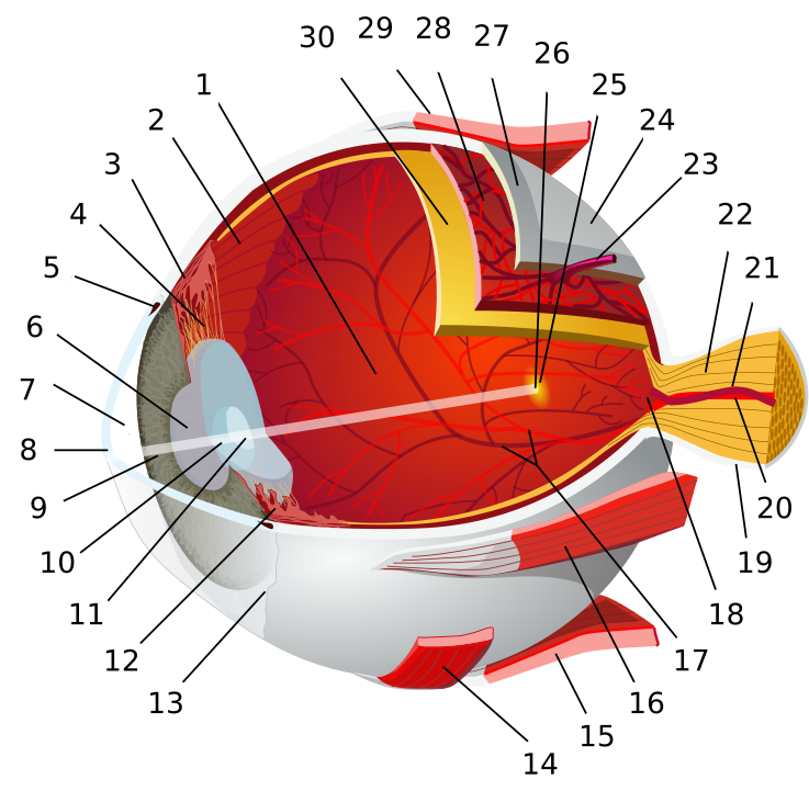
1:posterior segment of eyeball 2:ora serrata 3:ciliary muscle 4:ciliary zonules 5:canal of Schlemm 6:pupil 7:anterior chamber 8:cornea 9:iris 10:lens cortex 11:lens nucleus 12:ciliary process 13:conjunctiva 14:inferior oblique muscle 15:inferior rectus muscle 16:medial rectus muscle 17:retinal arteries and veins 18:optic disc 19:dura mater 20:central retinal artery 21:central retinal vein 22:optic nerve 23:vorticose vein 24:bulbar sheath 25:macula 26:fovea 27:sclera 28:choroid 29:superior rectus muscle 30:retina
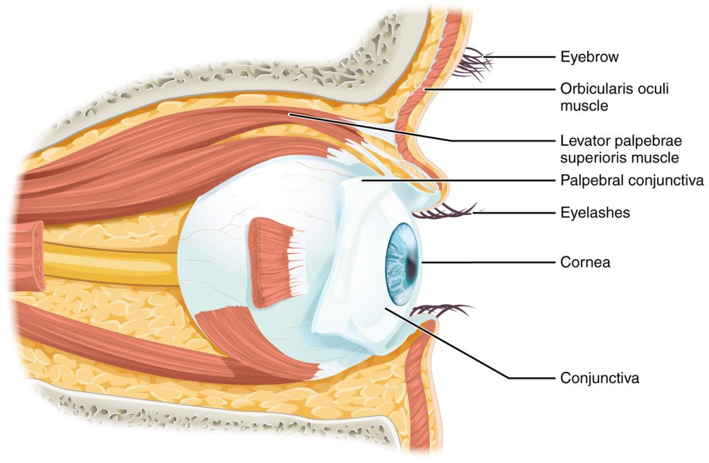
Movement of the eye within the orbit is accomplished by the contraction of six extraocular muscles that originate from the bones of the orbit and insert into the surface of the eyeball. Four of the muscles are arranged at the cardinal points around the eye and are named for those locations. They are the superior rectus, medial rectus, inferior rectus, and lateral rectus. When each of these muscles contract, the eye moves toward the contracting muscle. For example, when the superior rectus contracts, the eye rotates to look up. The superior oblique originates at the posterior orbit, near the origin of the four rectus muscles. However, the tendon of the oblique muscles threads through a pulley-like piece of cartilage known as the trochlea. The tendon inserts obliquely into the superior surface of the eye. The angle of the tendon through the trochlea means that contraction of the superior oblique rotates the eye medially. The inferior oblique muscle originates from the floor of the orbit and inserts into the inferolateral surface of the eye. When it contracts, it laterally rotates the eye, in opposition to the superior oblique. Rotation of the eye by the two oblique muscles is necessary because the eye is not perfectly aligned on the sagittal plane. When the eye looks up or down, the eye must also rotate slightly to compensate for the superior rectus pulling at approximately a 20-degree angle, rather than straight up. The same is true for the inferior rectus, which is compensated by contraction of the inferior oblique. A seventh muscle in the orbit is the levator palpebrae superioris, which is responsible for elevating and retracting the upper eyelid, a movement that usually occurs in concert with elevation of the eye by the superior rectus. The extraocular muscles are innervated by three cranial nerves. The lateral rectus, which causes abduction of the eye, is innervated by the abducens nerve. The superior oblique is innervated by the trochlear nerve. All of the other muscles are innervated by the oculomotor nerve, as is the levator palpebrae superioris. The motor nuclei of these cranial nerves connect to the brain stem, which coordinates eye movements.

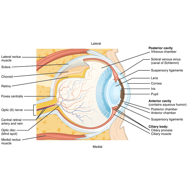
Identify the Structures of the Eye
Eyelids (Palpebrae) – Cover the eye
Meibomian glands – oily secretions – lubricate Medial and Lateral canthi
Conjunctiva – mucous membrane that covers the internal surface of the eyelids and much of the anterior eyeball
Eyeball
- Fibrous tunic – Dense irregular CT, avascular
- Sclera (white)
- Cornea – clear, refractory media of eyeball – bend light rays
- Vascular tunic, also called UVEA – carries blood to tissues of the eye
- Chroroid – posterior, brown, prevents light scattering in the eye
- Ciliary body – anterior, made mostly of ciliary muscle to control the shape of the lens
- Iris – most anterior portion – consist of muscle fibers around the pupil
- Sensory tunic b- Consist of
- Retina – contains photoreceptors
- Rods – shades of gray
- Cones – color
- Optic Nerve
- Lens – Divides the eyeball into
- Anterior Chamber – aqueous fluid = aqueous humors
- Posterior Chamber – viscous fluid = vitreous humor
Accessory structures
- Lacrimal apparatus
- Lacrimal glands
- Ducts – drain tears
Extraocular muscles – move eyeball
- Superior Rectus
- Inferior Rectus
- Medial Rectus
- Lateral Rectus
- Inferior Oblique
- Superior Oblique
- Innervation of the extraocular muscle of the eye
Ear
Hearing, or audition, is the transduction of sound waves into a neural signal that is made possible by the structures of the ear. The large, fleshy structure on the lateral aspect of the head is known as the auricle. Some sources will also refer to this structure as the pinna, though that term is more appropriate for a structure that can be moved, such as the external ear of a cat. The C-shaped curves of the auricle direct sound waves toward the auditory canal. The canal enters the skull through the external auditory meatus of the temporal bone. At the end of the auditory canal is the tympanic membrane, or ear drum, which vibrates after it is struck by sound waves. The auricle, ear canal, and tympanic membrane are often referred to as the external ear. The middle ear consists of a space spanned by three small bones called the ossicles. The three ossicles are the malleus, incus, and stapes, which are Latin names that roughly translate to hammer, anvil, and stirrup. The malleus is attached to the tympanic membrane and articulates with the incus. The incus, in turn, articulates with the stapes. The stapes is then attached to the inner ear, where the sound waves will be transduced into a neural signal. The middle ear is connected to the pharynx through the Eustachian tube, which helps equilibrate air pressure across the tympanic membrane. The tube is normally closed but will pop open when the muscles of the pharynx contract during swallowing or yawning.
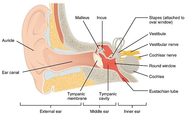
The inner ear is often described as a bony labyrinth, as it is composed of a series of canals embedded within the temporal bone. It has two separate regions, the cochlea and the vestibule, which are responsible for hearing and balance, respectively. The neural signals from these two regions are relayed to the brain stem through separate fiber bundles. However, these two distinct bundles travel together from the inner ear to the brain stem as the vestibulocochlear nerve. Sound is transduced into neural signals within the cochlear region of the inner ear, which contains the sensory neurons of the spiral ganglia. These ganglia are located within the spiral-shaped cochlea of the inner ear. The cochlea is attached to the stapes through the oval window.
The oval window is located at the beginning of a fluid-filled tube within the cochlea called the scala vestibuli. The scala vestibuli extends from the oval window, traveling above the cochlear duct, which is the central cavity of the cochlea that contains the sound-transducing neurons. At the uppermost tip of the cochlea, the scala vestibuli curves over the top of the cochlear duct. The fluid-filled tube, now called the scala tympani, returns to the base of the cochlea, this time traveling under the cochlear duct. The scala tympani ends at the round window, which is covered by a membrane that contains the fluid within the scala. As vibrations of the ossicles travel through the oval window, the fluid of the scala vestibuli and scala tympani moves in a wave-like motion. The frequency of the fluid waves match the frequencies of the sound waves. The membrane covering the round window will bulge out or pucker in with the movement of the fluid within the scala tympani.
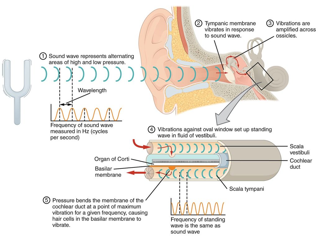
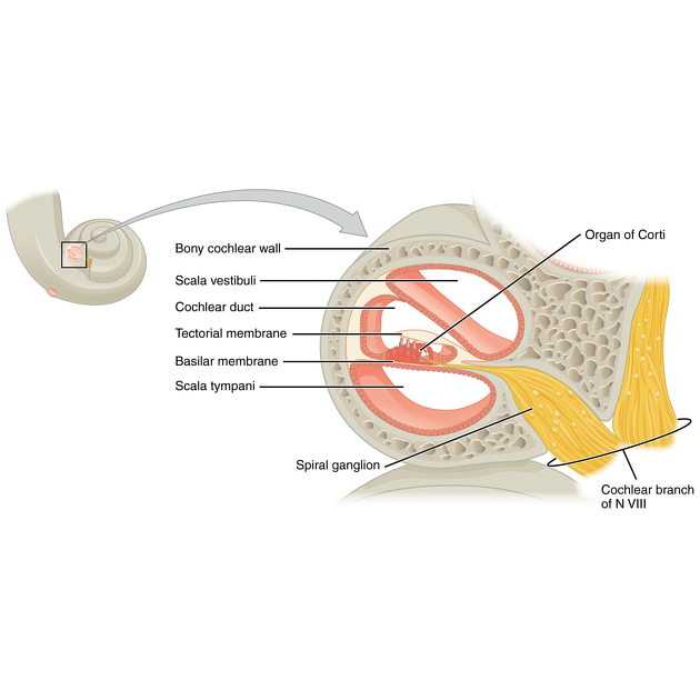
A cross-sectional view of the cochlea shows that the scala vestibuli and scala tympani run along both sides of the cochlear duct. The cochlear duct contains several organs of Corti, which tranduce the wave motion of the two scala into neural signals. The organs of Corti lie on top of the basilar membrane, which is the side of the cochlear duct located between the organs of Corti and the scala tympani. As the fluid waves move through the scala vestibuli and scala tympani, the basilar membrane moves at a specific spot, depending on the frequency of the waves. Higher frequency waves move the region of the basilar membrane that is close to the base of the cochlea. Lower frequency waves move the region of the basilar membrane that is near the tip of the cochlea.
Procedures
Plastic Anatomy – Identify structures.
- Review the muscles of the eye & ear in the images and tables provided using anatomical models available in the lab.
- Examine and label the eye & ear model and using images provided and anatomical models in the lab.
- Review the movement and action of each eye muscle using your eyes and your lab partners.
- Perform a Dissection of Cow eyeball – Watch the following video before coming to the lab.
You will need: lab coat, eye protection, dissection kit.
Media Attributions
- Eye diagram no circles border © By Chabacano - References: [1] [2] [3] among others, CC BY-SA 3.0, https://commons.wikimedia.org/w/index.php?curid=1759001 is licensed under a CC BY-SA (Attribution ShareAlike) license
- Eye in the Orbit © Wikipedia is licensed under a CC BY-SA (Attribution ShareAlike) license
- Extraocular muscles © Wikipedia is licensed under a CC BY-SA (Attribution ShareAlike) license
- Structure of the Eye © Radiopedia is licensed under a CC BY-SA (Attribution ShareAlike) license
- Structures of the ear © By OpenStax - https://cnx.org/contents/FPtK1zmh@8.25:fEI3C8Ot@10/Preface, CC BY 4.0, https://commons.wikimedia.org/w/index.php?curid=30147991 is licensed under a CC BY-SA (Attribution ShareAlike) license
- Sound Waves and the Ear © Wikipedia is licensed under a CC BY-SA (Attribution ShareAlike) license
- Cross Section of the Cochlea © Radiopedia is licensed under a CC BY-SA (Attribution ShareAlike) license

