The Cell
Pre-laboratory Activity – The Animal cell
Before you come to the lab, you should prepare yourself for the weekly lab activity, read the corresponding chapter in your textbook and complete the pre-lab activity.
Part 1 – Definitions
- Define the term cell

- Define the term cytology

- Define plasma membrane, cytoplasm, cytosol, nucleus and nucleolus

Part 2 – Drawing Activity

- Draw an animal cell.
- List the components of the cell using different colors.
- Color the structures of an animal cell using the color scheme you developed. Use a different color for each of the cell components if possible.
Laboratory Activity: The Animal Cell: Structure and Function
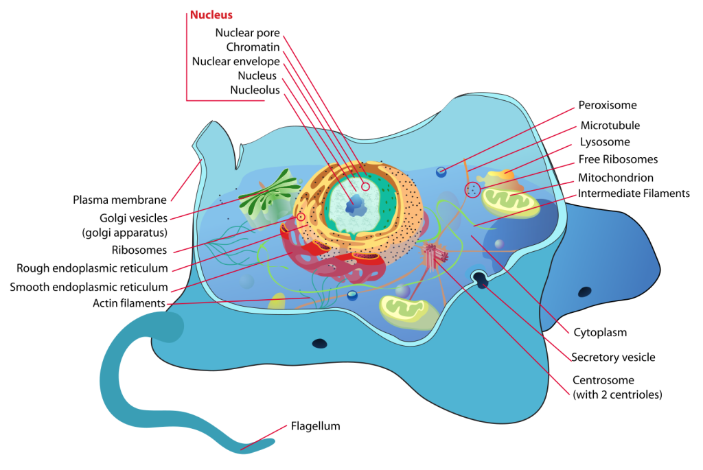
Introduction
The term cell comes from the Latin word “cella”, meaning “small room”. Cells were discovered by Robert Hooke in 1665, who named them for their resemblance to cells inhabited by Christian monks in a monastery.
The cells is a small container, it is the basic structural, functional, and biological unit of all known living organisms. A “cell” is the smallest unit of life known to man. Cells consist of a semi solid fluid, called cytoplasm and enclosed within a plasma membrane, which contains of biomolecules such as proteins, carbohydrates, lipids and nucleic acids as well as ions. Humans contain about 30 trillion (3013) cells. Cells size varies between 1 and 100 micrometers, therefore they are only visible only under a microscope.
The “Cell theory” was developed in 1839 by Matthias Jakob Schleiden and Theodor Schwann, it states that all organisms are composed of one or more cells, that cells are the fundamental unit of structure and function in all living organisms, and that all cells come from pre-existing cells.
It is believed that cells emerged on Earth at least 3.5 billion years ago, a common ancestor that has given rise to all organisms we have on earth.
Components of the Cell
Animal cells have a number of organelles and structures that perform specific functions inside the cell and allow them to communicate with other cells, to move or move particulate away from their surface.
Cells have evolved to perform different functions therefore they do not always all the organelles or structures we show here, but we will introduce you to most of the organelles present in animal cells.
PLASMA MEMBRANE
The plasma membrane is a membrane that surrounds an animal cell, it is the boundary between the inside and the outside of the cell. The plasma membrane is made of a double layer of phospholipids. Other compounds such as proteins and carbohydrates may be embedded into the plasma membrane and are responsible to receive cellular signals from other cells, and to allow passage of substances in and out of the cell by creating channels through the membrane.
NUCLEUS
The nucleus is an organelle found in eukaryotic cells. It is fully enclosed by nuclear membrane and it contains the majority of the cell’s genetic material.
The nucleus consists of a nuclear envelope, chromatin, and a nucleolus. The nuclear envelope is composed of a set of double membranes with numerous pores (nuclear pores) to allow substances to move in and out of the nucleus. Chromatin contains the majority of a cell’s DNA. It condenses to form chromosomes during cell division. The nucleolus is the center core of the nucleus. Nucleoli are made of proteins, DNA and RNA and it is responsible for ribosome biogenesis. The nucleolus makes ribosomal subunits from proteins and ribosomal RNA (rRNA). It then sends the subunits out to cytoplasm where they combine into complete ribosome units.
The main function of the cell nucleus is to control gene expression and mediate the replication of DNA during the cell cycle.
CYTOPLASM
The cytoplasm is the internal area of an animal cell. It consists of a jelly-like substance called ‘cytosol’ and allows organelles and cellular substances to move around the cell as needed.
ENDOPLASMIC RETICULUM (ER)
The endoplasmic reticulum is a network of membranes that originate from the nuclear envelope. The membranes are important for many cellular processes such as protein production and the metabolism of lipids and carbohydrates
The endoplasmic reticulum includes; the smooth ER and the rough ER. The smooth ER is a smooth membrane and has no ribosomes, whereas the rough ER has ribosomes that are used to produce proteins.
MITOCHONDRIA
They are the site of cellular respiration – the process that breaks down sugars and other compounds into cellular energy (ATP). It is in the mitochondria where oxygen is used as a final electron acceptor in the respiratory chain to generate many molecules of ATP.
GOLGI APPARATUS
The Golgi apparatus (or Golgi body) is a set of membranes found within the cell but is not attached to the nucleus of the cell. It serves many important functions including post-translational modification of proteins and lipids and transporting cellular substances inside and outside the cell.
RIBOSOMES
Ribosomes are involved in proteins synthesis. They can be either attached to the endoplasmic reticulum or floating freely in the cell’s cytoplasm. They are formed by two subunits of ribosomal RNA.
PEROXISOMES
S organelles involved in the digestion of compounds such as fats, amino acids, and sugars. They also produce hydrogen peroxide.
LYSOSOMES
Lysosomes are essentially waste disposal units. They contain a range of enzymes that digest molecules such as lipids, carbohydrates, and proteins.
CENTROSOMES
Centrosomes are involved in cell division and the production of flagella and cilia. They consist of two centrioles that are the main hub for a cell’s microtubules. As the nuclear envelope breaks down during cell division, microtubules interact with the cell’s chromosomes and prepares them for cellular division.
VILLI
Villi are hair-like extensions of the plasma membrane. They are important for absorption of substances from their surrounding environment because they essentially increase the surface area of a cell.
FLAGELLA
Flagella are appendages that are involved in movement. Flagella (plural of flagellum) provide the mechanical ability for cells to move under their own power.
A flagellum is a long, thin extension of the plasma membrane and is driven by a cellular engine made from proteins. Flagella are particularly important for certain animal cells. In humans the only cell that contains flagella is the sperm cells,
DIFFERENT TYPES OF ANIMAL CELLS
There are many different types of animal cells. I will mention only a few here.
SKIN CELLS
The skin cells of animals mostly consist of keratinocytes and melanocytes
Melanocytes produce a compound called ‘melanin’ which gives skin its color.
MUSCLE CELLS
Also called myocytes, muscle fibers or muscle cells are long tubular cells responsible for moving an organism’s limbs and organs. Muscle cells can be either skeletal muscle cells, cardiac muscle cells or smooth muscle cells.
BLOOD CELLS
Blood cells: red blood cells and white blood cells. Red blood cells make up around 99.9% of all blood cells and are responsible for delivering oxygen from the lungs to the rest of the body. Red blood cells do not have a nucleus. White blood cells are a vital part of an animal’s immune system and help to battle infections by killing off damaging bacteria or damaged/infected cells.
NERVE CELLS
There are two types of nerve cells, neurons and neuroglia. There are many more neuroglia than neurons. Neuroglia are there to support, protect and nourish neurons. The human brain alone has ~ 100 billion neuron cells. They allow communication between cells. They contain a body, many dendrites and one axons. Dendrites and axons are extensions from the cell that receive and transport signals to and from the cell, respectively.
FAT CELLS
Fat cells, also known as adipocytes. They are used to store fat as energy reserves.
Materials
- 24-color pencil set (students should bring their own set of color pencils)
- Black sharpie
- Labeling tape
- Individual slides:
- Simple squamous epithelium (cheek cells)
- Columnar epithelium (isolated)
- Smooth muscle (teased)
- Skeletal muscle (teased)
- Human blood (smear)
- Fish blastocyst slide (mitosis)
- Demonstration slides of organelles with special stains/treatments (these slides will be set up as demo stations at the instructor’s bench).
- Sickle cell anemia
- Cardiac muscle (Intercalated disk stain)
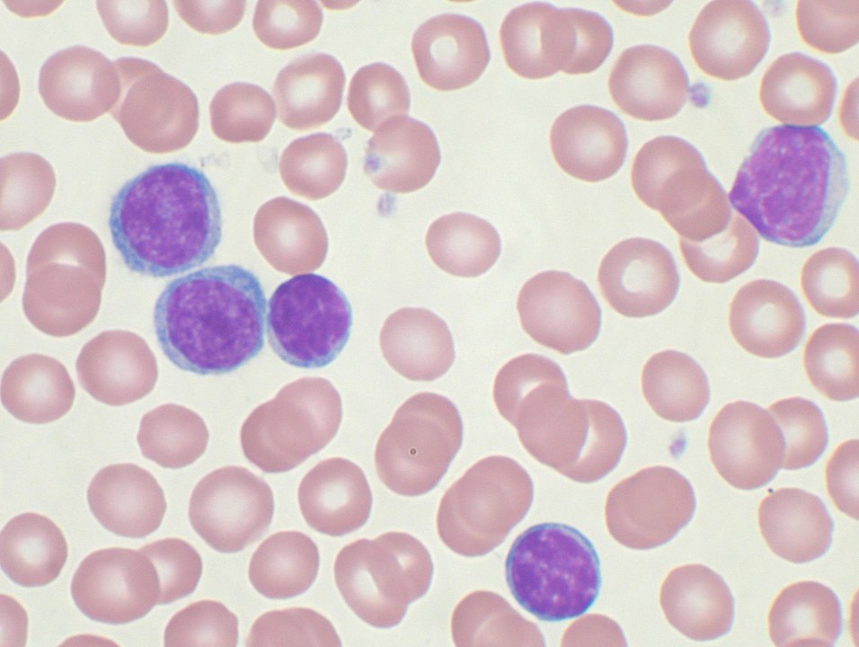
- Lymphocytic leukemia
- Anatomical models
- 3-D anatomical model of an animal cell (with key).
- 3-D model of mitosis (with key).
Note: the type of slides may vary depending on what is available in a laboratory.
Review the previous lab, the microscope as you will be using the microscope for this activity and for many others during the semester.
You will also need to review your textbook and any material your instructor has assigned as supplemental reading, as well as posted videos and animations related to the animal cell, the cell cycle and mitosis.
Objectives
- Define the terms cell and cytology
- List and identify the parts of an animal cell
- List the functions of main components of an animal cell
Laboratory Activity – The animal cell and its components
A. Identify the following parts of the cell using a 3-D model of an animal cell
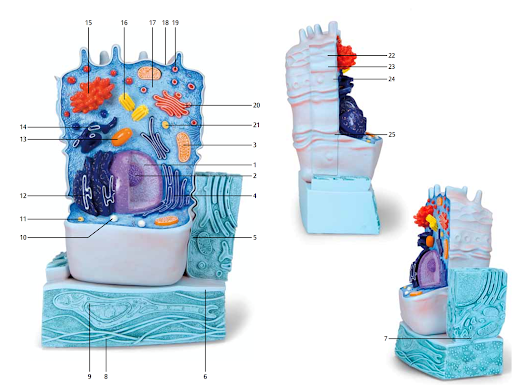
B. Cell types – observe and draw the following cells types.
| Number | Part of the cell or organelle |
| 1 | |
| 2 | |
| 3 (same as #13, l.s.) | |
| 4 (same as #14, l.s.) | |
| 5 | |
| 6 | |
| 7 | |
| 8 | |
| 9 | |
| 10 | |
| 11 | |
| 12 | |
| 13 | |
| 14 | |
| 15 | |
| 16 | |
| 17 | |
| 18 | |
| 19 | |
| 20 (same as #15, l.s.) | |
| 21 | |
| 22 | |
| 23 | |
| 24 | |
| 25 |
Instructions: Using the microscope observe slides of simple columnar epithelium, skeletal muscle and a human blood smear.
Use scanning objective to locate the samples, then switch to low or high power to be able to see the cell morphology.
If these types of cells are not available use what your instructor provides for you.
B1. Simple columnar epithelium
Observe tall rectangular cells with the nuclei oriented toward the base of the cells. Then draw what you see in the space provided. Make sure to write down the total magnification used.
B2. Skeletal Muscle Fiber
Observe long cells with a banding pattern and the nuclei oriented toward periphery. Then draw what you see in the space provided. Make sure to write down the total magnification used.


B3. Human blood smear
- Observe a human blood smear under the microscope. First use the scanning objective to locate the sample, then switch to higher objectives.
- To see the shape of the cells you need to use the high power objective (40X).
- You will see the formed elements of the blood, predominantly red blood cells (background) and a few white blood cells (cells with transparent cytoplasm and a dark purple nucleus). You may also see very small fragments of cells called platelets (few).
- Then draw what you see in the space provided. Make sure to write down the total magnification used.

C. Demonstration slides (special stains/treatments or disease states)
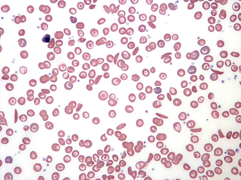
- Sickle cell anemia
- Cardiac muscle (Intercalated disk stain)
- Lymphocytic leukemia
Observe the difference in cell morphology on the slides set as a demo on the instructor’s bench.
Note: The type of slide may vary depending on what is available in the lab.
D. Mitosis
D1. Fish Blastocyst
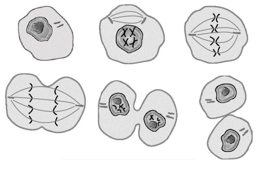
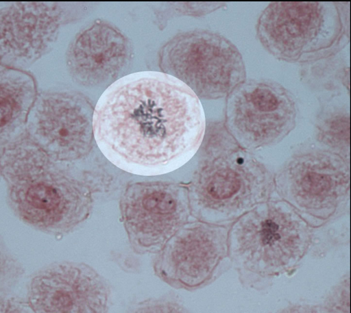
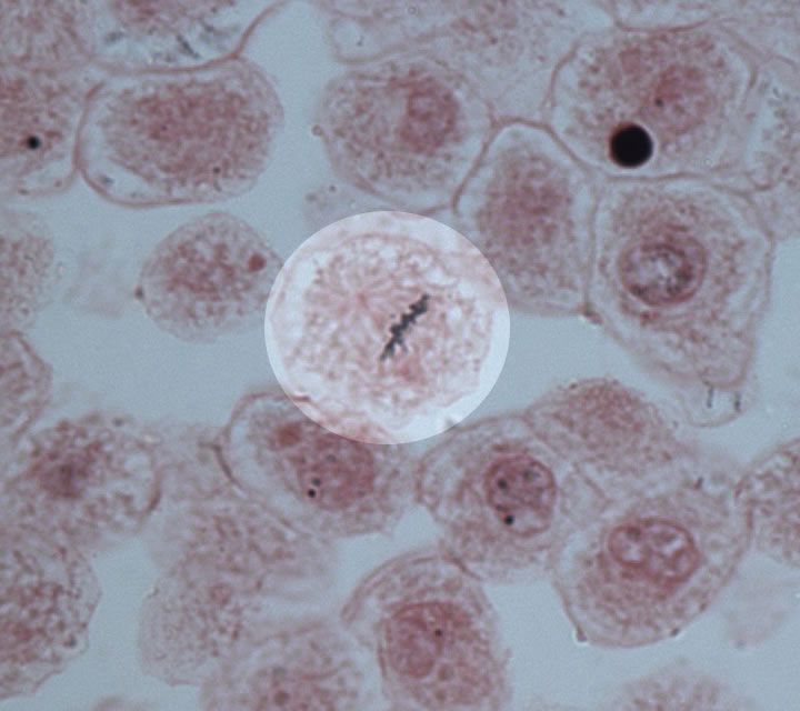
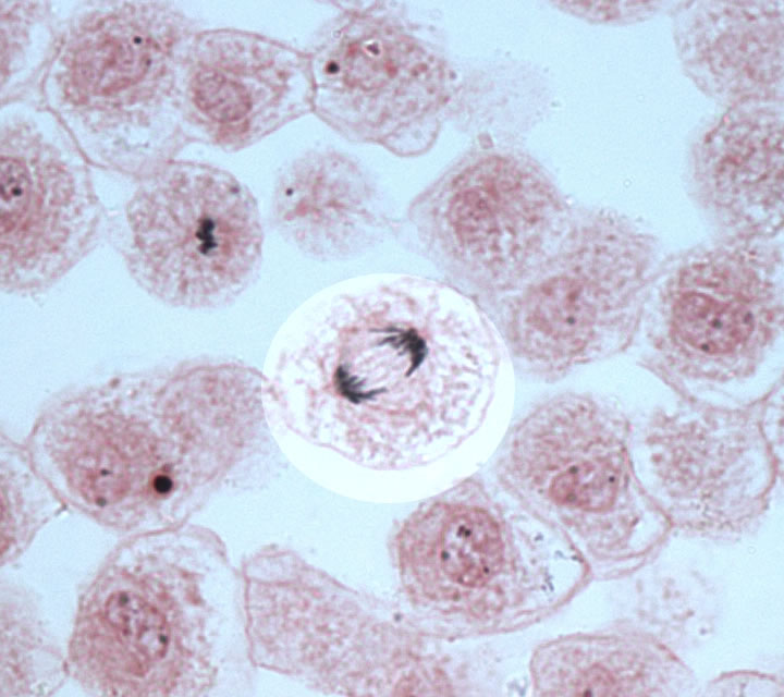
Obtain a microscope slide of Fish Blastocyst and try to find and identify the different stages of mitosis. Then draw what you see in the space provided. Make sure to write down the total magnification used.


D2. Label the stages of Mitosis using the images shown here
For 100% online labs use the following links:
- Simple columnar epithelium – UM Slide # 176: Colon, H&E, 40X (simple columnar epithelium)
- Skeletal Muscle Fiber – UM Slide #058L – Skeletal muscle, longitudinal section, H&E, 20X and 40X (banding pattern A I Z H [60787 x 18392, 34840 x 9034, 64339 x 17968])
- Human blood smear – UM Slide #086: Human blood smear, Giemsa stain, 86X scan from hematopathology normals collection (this slide contains 3 basophil cells)
- Sickle cell anemia
- Cardiac muscle (Intercalated disk stain) – UM Slide #098-1: Heart, ventricle, H&E, 40X (cardiac muscle, intercalated discs)
- Lymphocytic leukemia
- Fish Blastocyst
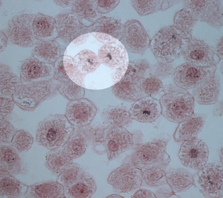
Whitefish embryos are commonly used in the study of mitosis due to their rapid cell division and relatively large and transparent cells. Here are some images of mitosis in white fish blastocysts.
Visit the Biology Corner
Media Attributions
- Animal cell structure © Wikipedia is licensed under a CC BY (Attribution) license
- Figure 2. 3D animal cell for lab activity © Maria Carles adapted by 3B Scientific is licensed under a CC BY-SA (Attribution ShareAlike) license
- Blank circle © Maria Carles is licensed under a CC BY-SA (Attribution ShareAlike) license
- Stages of Mitosis © Learn 4 Your Life is licensed under a All Rights Reserved license
- Sickle Cell Blood Smear © Wikipedia is licensed under a CC0 (Creative Commons Zero) license
- Lymphocytic leukemia © By VashiDonsk at the English Wikipedia, CC BY-SA 3.0, https://commons.wikimedia.org/w/index.php?curid=2111159 is licensed under a CC0 (Creative Commons Zero) license
- Prophase © The Biology Corner is licensed under a CC BY-NC-SA (Attribution NonCommercial ShareAlike) license
- Metaphase © The Biology Corner is licensed under a CC BY-NC-SA (Attribution NonCommercial ShareAlike) license
- Anaphase © The Biology Corner is licensed under a CC BY-NC-SA (Attribution NonCommercial ShareAlike) license
- Telophase © The Biology Corner is licensed under a CC BY-NC-SA (Attribution NonCommercial ShareAlike) license

