The Skeletal System Anatomy
The Skeletal System Laboratory Activity
Objectives
- Define and differentiate between the axial skeleton and the appendicular skeleton
- Identify the major bones and markings of the axial skeleton
- Identify the major bones and markings of the appendicular skeleton
- Build a skeleton using disarticulated bones
Introduction
This laboratory activity will introduce the students to the bones and structures that form the human skeleton. The skeleton is the internal framework of the human body. It is composed of 206 bones in the adult body.
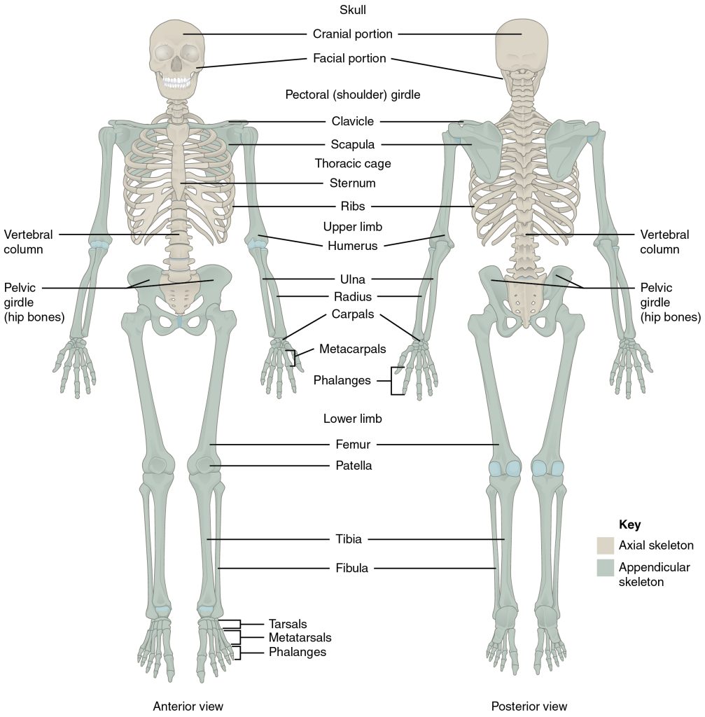
The human skeleton can be divided into two portions: the axial skeleton and the appendicular skeleton.
The axial skeleton is formed by the rib cage (vertebral column, ribs, and sternum), the skull, mandible, hyoid bone and the three ossicles (inside the ear).
The appendicular skeleton, which is attached to the axial skeleton, is formed by the shoulder girdle, the pelvic girdle and the bones of the upper and lower limbs.
The human skeleton performs six major functions; support, movement, protection, production of blood cells, storage of minerals, and endocrine regulation.
Laboratory Activity
In this lab, you will learn to identify the bones of the adult human skeleton, its processes, orientation and how they articulate with each other using sets of disarticulated and articulated skeletons.
Part 1 – Identify the bones of the human skeleton. Label the bones using the images shown here.
- Axial Skeleton – Identify bones and structures of the skull
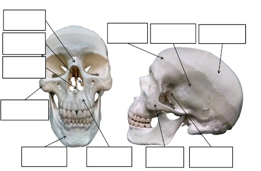
Label the following:
Zygomatic bones; Lacrimal bones, Occipital bone; Coronal suture; Sagittal suture; Superior orbital notch/foramen; Inferior orbital foramen; Glabella; Superior orbital fissure; Inferior orbital fissure.
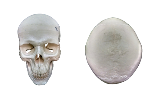
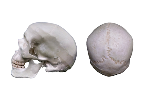
Label the following:
Occipital bone; Parietal bone; Temporal bone; Sphenoid bone; Maxilla; Mandible; Zygomatic bone; External acoustic meatus; Mastoid process; Styloid process; Lambdoid suture; Squamous suture; Zygomatic process of the temporal bone; Temporal process of the zygomatic bone; Wormian (or sutural) bones.
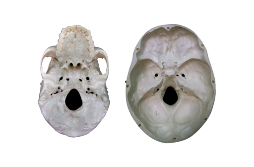
Label the following:
Zygomatic arch; Lesser wing of the sphenoid; Sella turcica; Foramen ovale; Foramen spinosum; Foramen lacerum; Jugular foramen; Carotid canal; Foramen magnum; Internal acoustic meatus; Occipital condyle; Mandibular fossa; Ethmoid bone; Crista galli; Optic canal; Anterior cranial fossa; Middle cranial fossa; Posterior cranial fossa.
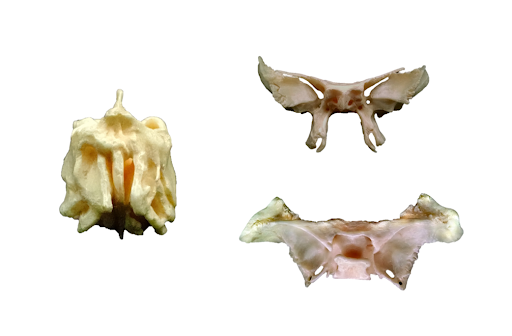
Label the following:
Ethmoid bone: Perpendicular plate; Crista galli; Lateral masses.
Sphenoid bone: Lesser wing; Greater wing; Sella turcica; Pterygoid plates; Hypophyseal fossa.
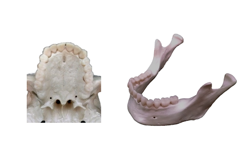
Label the following:
Maxilla; Palatine bone; Pterygoid plates; Vomer, Greater palatine foramen, Incisive foramen
Mandible; Condylar process; Coronoid process; Mental foramen; Body of mandible; Ramus of mandible; Mandibular notch.
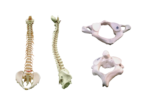
Label the following:
Cervical vertebrae; Cervical curvature; Thoracic vertebrae; Thoracic curvature; lumbar vertebrae; Lumbar curvature; Sacrum; Coccyx; 7; 12; 5.
Atlas; Axis; Dens; C1; C2; Spinous process; Superior articular facet.
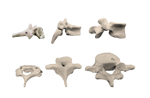
Label the following:
Cervical vertebrae; Thoracic vertebrae; Lumbar vertebrae; Transverse foramen; Superior articular process; Inferior articular process; Costal demifacet; Vertebral foramen; Body; Spine; Arch; Transverse process; Transverse facet
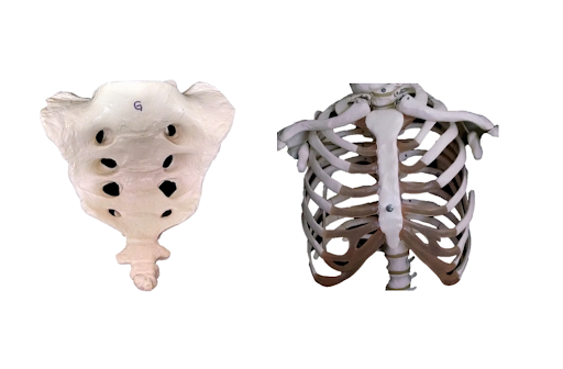
Label the following:
Sacrum: Sacral foramen; Ala (of the sacrum); Auricular surface.
Coccyx.
Sternum: Manubrium; Body; Xiphoid process; Jugular notch; Sternal angle.
True ribs; False ribs; Floating ribs; Costal cartilages.
- B. Appendicular Skeleton – Identify bones and structures of the appendicular skeleton
Label the following:
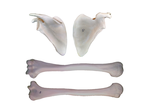
Label the following:
Scapula: Spine; Coracoid process; Acromion; Supra-spinous fossa; Infra-spinous fossa; Sub-scapular fossa; Glenoid cavity
Humerus: Head; Neck; (surgical and anatomical); Trochlea; Capitulum; Olecranon fossa; Coronoid fossa; Radial fossa; Medial epicondyle; Lateral epicondyle; Greater tubercle; Lesser tubercle
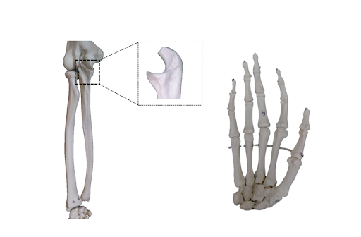
Label the following:
Radius: Styloid process; Radial head; Radial tuberosity.
Ulna: Olecranon; Trochlear notch; Coronoid process; Styloid process;
Carpals: Scaphoid; Lunate; Triquetral; Pisiform; Trapezium; Trapezoid; Capitate; Hamate.
Metacarpals
Phalanges: proximal; middle and distal
Pollex
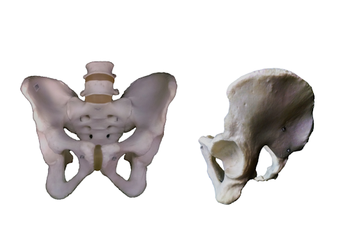
Label the following:
Ilium; Ischium; Pubis.
Obturator foramen; Acetabulum; Iliac crest; Anterior superior iliac spine; Anterior inferior iliac spine; Posterior superior iliac spine; Posterior inferior iliac spine; Pubis symphisis; Greater sciatic notch; Ischial spine; Sacrum; (male or female pelvis).
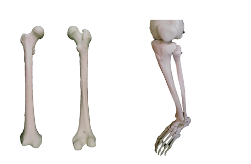
Label the following:
Femur: Head; Neck; Greater trochanter; Lesser trochanter; Medial condyle; Lateral condyle; Medial epicondyle; Lateral epicondyle; Linea aspera.
Tibia: Medial malleolus; Tibial tuberosity.
Fibula: Lateral malleolus.
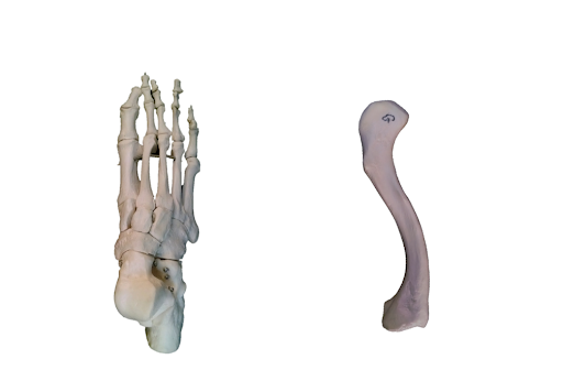
Label the following:
Tarsals: Talus; Calcaneus; Cuboid; Navicular; Medial,intermediate & lateral cuneiforms;
Metatarsals
Phalanges: proximal; middle; distal.
Hallux
Clavicle: sternal end, acromial end
Appendix A – List of bone and its structures
This is the list of bones and structures that you must be able to identify after performing this lab activity. You should use the articulated and disarticulated skeletons available in the laboratory to practice identifying all structures listed here. It is recommended to work in groups of two and to test each other as you practice the identification of all structures listed here.
Axial Skeleton
Human Skull
Cranial Bones (bones that form the cranial cavity)
- Bones forming the calvarium (skull cap)
- Frontal bone (1)
- Supraorbital margins (ridges)
- Supraorbital foramina (notches)
- Parietal bone (2)
- Occipital bone (1)
- Foramen Magnum
- Occipital Condyles
- External occipital protuberance
- Frontal bone (1)
Bones forming the Floor of the cranial cavity
- Frontal (1)
- Frontal sinus
- ETHMOID BONE (1)
- Cribriform plate
- Crista galli
- Olfactory foramina (passage for olfactory nerves)
- Perpendicular plate (anterior and superior part of nasal septum, articulates with vomer)
- Superior nasal conchae (turbinate bones)
- Middle nasal conchae (turbinate bones)
- Ethmoid sinus (air cells)
- SPHENOID BONE (1)
- Lesser wings
- Greater wings
- Sella turcica
- Sphenoid sinus
- Optic foramina
- Temporal bone (2)
- Squamous portion – squamous suture, zygomatic process of the temporal bone (part of zygomatic arch), mandibular fossa
- Tympanic portion – external auditory meatus, styloid process and stylomastoid foramen
- Mastoid portion – mastoid process and mastoid air cells
- Petrous portion – internal auditory meatus, carotid foramen and canal, jugular foramen
- Occipital bone (1)
Other structures of the cranial floor
- Anterior, Middle and Posterior Fossae
Facial bones (bones of the face)
- Mandible (1)
- Mental Protuberance
- Alveolar process
- Rami of mandible
- Angle of mandible
- Body of mandible
- Mandibular condyles
- Mental foramina
- Maxilla (2):
- Maxillary sinus
- Alveolar process
- Palatine process (2/3 of hard palate)
- Palatine bone (2) (forms posterior 1/3 of hard palate)
- Zygomatic bone – ZYGOMA (2)
- Temporal process of the zygomatic bone
- Lacrimal bone (2)
- Lacrima fossae
- Nasal bone, also called Nasalis (2), they form the bridge of the nose
- Inferior nasal conchae (2)
- Vomer (1), forms inferior and posterior nasal septum, articulates with perpendicular plate of the ethmoid bone
BONES OF THE ORBIT
- Frontal
- Zygomatic
- Maxillary
- Sphenoid
- Ethmoid
- Palatine
- Lacrimal
OSSICLES (three bones in middle ear)
- Malleus
- Incus
- Stapes
- Hyoid bone (supports the tongue)
VERTEBRAL COLUMN
- Cervical vertebrae (7)
- C1 = Atlas
- C2 = Axis
- Dens or odontoid process
- Thoracic vertebrae (12)
- Lumbar vertebrae (5).
- Sacrum (5, fused sacral vertebrae)
- Coccyx (4-6, fused coccygeal vertebrae) vestigial tail
- VERTEBRA – Structures
- Body of Vertebra
- Spinous process
- Transverse process
- Vertebral foramen
- Superior articular facet
- Inferior articular facet
- Intervertebral foramen
THORACIC CAGE
- STERNUM
- Manubrium
- Body of sternum
- Xiphoid process
- RIBS
- Ribs 1-7 (true ribs)
- Ribs 8-12 (false ribs)
- Ribs 11-12 (floating ribs)
- Ribs – Structures:
- Head
- Neck
- Tubercle
- Body
- Costal groove
- Sternal end
SUTURES
- Frontal or Coronal
- Sagittal
- Lambdoidal
- Squamous
- Sutural bones or Wormian bones
Fetal Skull
- Anterior fontanelle
- Posterior fontanelle
- Anterolateral fontanelle
- Posterolateral fontanelle
Appendicular Skeleton
GIRDLES AND LIMBS
PECTORAL GIRDLE
- Clavicle
- Acromial end
- Sternal End
- Scapula
- Spine
- Acromion
- Glenoid cavity
- Coracoid process
- Supraspinous fossae
- Infraspinous fossae
- Subscapular fossae
- Axillary border
- Vertebral border
- Superior border
- Inferior border
UPPER LIMB (Humerus, radius, ulna, carpals, metacarpals, phalanges)
- Humerus (brachium – arm)
- Proximal end:
- Head
- Anatomical neck
- Surgical neck
- Greater tubercle
- Lesser tubercle
- Shaft:
- Deltoid tuberosity
- Distal end:
- Capitulum
- Trochlea
- Lateral epicondyle
- Medial epicondyle
- Olecranon fossa
- Radius (antebrachium – forearm)
- Proximal end:
- Head
- Radial tuberosity
- Distal end:
- Styloid process
- Ulnar notch
- Ulna (antebrachium – forearm):
- Proximal end:
- Olecranon
- Radial notch
- Trochlear notch
- Distal end:
- Head
- Styloid process
- Wrist (carpus) – carpal bones (8)
- Scaphoid, lunate, triquetrum, pisiform, trapezium, trapezoid, capitate, hamate
- Hand (Manus – 19 bones):
- Metacarpus
- Metacarpal bones ( I – V)
- Phalanges ( I – V)
- Proximal, middle, distal phalanx
PELVIC GIRDLE (hip girdle, 2 innominate bones with sacrum)
- 2 coxal bones, also called innominate bones and os coxae.
- Ilium:
- Iliac crest
- Iliac fossa
- Greater sciatic notch
- Acetabulum (formed by the fusion of ilium, ischium and pubis)
- Ischium:
- Ischial spine
- Ischial tuberosity
- Lesser sciatic notch
- Acetabulum (formed by the fusion of ilium, ischium and pubis)
- Obturator foramen
- Pubis:
- Pubic symphysis
- Acetabulum (formed by the fusion of ilium, ischium and pubis)
- Obturator foramen
LOWER LIMB (femur, patella, tibia, fibula, tarsals, metatarsals, phalanges)
- Femur (thigh)
- Proximal end:
- Head
- Neck
- Greater trochanter
- Lesser trochanter
- Gluteal tuberosity
- Shaft:
- Linea aspera
- Distal end:
- Patellar surface
- Medial condyles
- Lateral condyles
- Medial epicondyles
- Lateral epicondyles
- Patella (kneecap)
- Base
- Apex
- Tibia (leg, lower leg)
- Proximal end:
- Medial condyles
- Lateral condyles
- Tibial tuberosity
- Shaft:
- Anterior crest
- Distal end:
- Medial malleolus
- Proximal end:
- FIBULA (leg)
- Proximal end:
- Head
- Distal end:
- Lateral malleolus
- Proximal end:
- ANKLE (tarsus)
- 7 tarsal bones: talus, calcaneus, navicular, medial, intermediate and lateral cuneiformns, cuboid
- Foot (pes – 19 bones)
- Metatarsus
- Metatarsal bones (I-V)
- Phalanges (I-V):
- Proximal, middle, distal phalanx
Media Attributions
- Skeleton from the Side © Wikipedia is licensed under a Public Domain license
- Axial skeleton © Wikipedia is licensed under a CC BY-SA (Attribution ShareAlike) license
- Label axial skeleton © Maria Carles is licensed under a CC BY-SA (Attribution ShareAlike) license
- Label Zygomatic bones 1 © Maria Carles is licensed under a CC BY-SA (Attribution ShareAlike) license
- Label Zygomatic bones 2 © Maria Carles is licensed under a CC BY-SA (Attribution ShareAlike) license
- Label Occipital bone © Maria Carles is licensed under a CC BY-SA (Attribution ShareAlike) license
- Label Zygomatic arch © Maria Carles is licensed under a CC BY-SA (Attribution ShareAlike) license
- Label Ethmoid bone © Maria Carles is licensed under a CC BY-SA (Attribution ShareAlike) license
- Label Maxilla © Maria Carles is licensed under a CC BY-SA (Attribution ShareAlike) license
- Label Cervical vertebrae © Maria Carles is licensed under a CC BY-SA (Attribution ShareAlike) license
- Label Cervical vertebrae 2 © Maria Carles is licensed under a CC BY-SA (Attribution ShareAlike) license
- Label Sacrum © Maria Carles is licensed under a CC BY-SA (Attribution ShareAlike) license
- Label Scapula © Maria Carles is licensed under a CC BY-SA (Attribution ShareAlike) license
- Label Radius © Maria Carles is licensed under a CC BY-SA (Attribution ShareAlike) license
- Label Ilium © Maria Carles is licensed under a CC BY-SA (Attribution ShareAlike) license
- Label Femur © Maria Carles is licensed under a CC BY-SA (Attribution ShareAlike) license

