The Nervous Tissue
Neuron Anatomy & Physiology
Introduction
The nervous system is one of the two major communicating systems of the body. It works together with the endocrine system to control, regulate, all system in the body to maintain homeostasis.
It is the center of all mental activity including thought, learning, and memory. Through its receptors, the nervous system keeps us in touch with our internal and external environments.
The nervous system is composed of organs, principally the brain, spinal cord, nerves, and ganglia. These, in turn, consist of various tissues, including nervous, epithelial, and connective tissue. Together these carry out the complex activities of the nervous system.
The activities of the nervous system can be grouped as three general overlapping functions; sensory, integrative and motor.
The nervous tissue is composed of two types of cells, neurons (functional units, capable of transmitting an action potential) and neuroglia that support and nourish the neurons, usually they are found in a ratio of 1:9 (neuron:neuroglia).
These cells are contained in the organs of the nervous system.
The Central Nervous System (CNS) is composed of the brain and the spinal cord, and the Peripheral Nervous System (PNS) is composed of all tissue located outside of the CNS, cranial and peripheral nerves.
Objectives:
- Distinguish functions of the neurons and neuroglia
- Identify the anatomical characteristics of a neuron
- Classify neurons based on structure and function
- Describe the structure of a nerve and the connective tissue coverings
Procedures
A. Gross Anatomy of a Neuron
Use a 3D model of a neuron (Somso, BS35) to identify the following structures:
- Perikaryon (nerve cell)
- Peripheral Nerve with sheaths
- Neurite cone
- Nucleus of nerve cell with nucleolus
- BS35: Endoplasmic reticulum, BS35-1 Nissl’s granules
- Neurofibrils
- Synaptic terminals
- Neuroaxon
- Schwan cells with nucleus
- Schwan sheath
- Linked Schwan cells at node of Ranvier
- Mitochondria
- Medullary myelin sheath
- Perineural sheath of connective tissue
- Mesaxon
- Dendrites
- Lysosome
- Neurotubules
- Golgi Apparatus
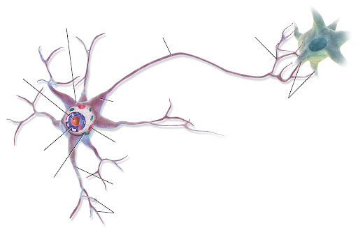
Use the following diagram to label and draw any missing structure of a neuronal network
- Cell body
- Nucleus
- Nissl bodies
- Mitochondria
- Axon Hilock
- Dendrites
- Axons
- Myelin sheath
- Node of Ranvier
- Internodes
- Synaptic terminals – Telodendria
- Pre-synaptic cell
- Post-synaptic cell
- Synaptic cleft

B. Microscopic Anatomy of a Neuron
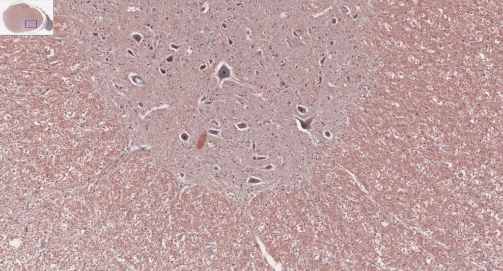
Use the slides or a virtual microscope (link provided) to draw and label the microscopic structures of a neuron:
- Motor neuron
- Spinal cord smear
Find/label the following structures:
- Motor neurons, cell body, nucleus, dendrites, axons and Nissl bodies (this may not be visible in all slides)
Virtual Microscopy, UM Virtual Microscope or QR code below.

- Slide: # 65-1 Spinal cord and dorsal root ganglion, H&E, 20X (white matter [pinkish], gray matter [grayish], dorsal horn, ventral horn, ventral horn cells [large motor neuron cell bodies], neuropil, dorsal root ganglion [at right], sensory neurons, capsular cells, sensory axon tracts).

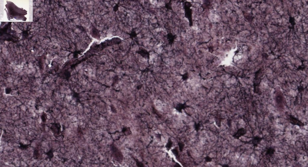
- Neuroglia cell bodies (Astrocytes), Slide # 13270, Astrocytes, Gold-staining

C. Neuron Classification
Draw, label and describe the function of the following types of neurons:
- Multipolar

- Bipolar

- Unipolar

D. Structure of a nerve
Using your textbook and/or the internet, draw and name the three connective tissue coverings of a nerve.
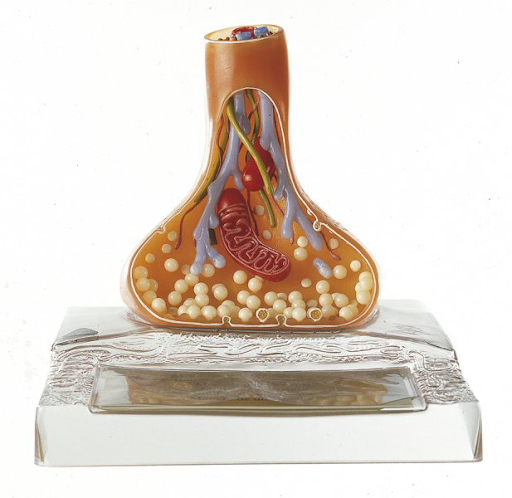
Identify & label the parts of a Synapse.

Media Attributions
- Neuronal network © Maria Carles is licensed under a CC BY-SA (Attribution ShareAlike) license
- QR code V Microscope © Maria Carles is licensed under a CC BY-SA (Attribution ShareAlike) license
- Spinal cord and dorsal root ganglion © Maria Carles adapted by Michigan Medical School is licensed under a CC BY-NC-SA (Attribution NonCommercial ShareAlike) license
- Astrocytes © Maria Carles is licensed under a CC BY-NC-SA (Attribution NonCommercial ShareAlike) license
- Label Synapse © Maria Carles is licensed under a CC BY-SA (Attribution ShareAlike) license

