The Cardiovascular System Anatomy
Cardiovascular System Laboratory Activity
The Heart and Associated Vessels
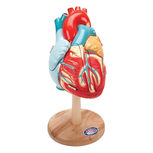
The heart is a muscular organ that pumps blood through the blood vessels of the circulatory system. The pumped blood carries oxygen and nutrients to the body, while carrying metabolic waste such as carbon dioxide to the lungs.
In humans, the heart is approximately the size of a closed fist and is located between the lungs, in the middle compartment of the chest, called the mediastinum. The heart is enclosed in a protective sac, the pericardium, which also contains a small amount of fluid. The wall of the heart is made up of three layers: epicardium, myocardium, and endocardium.
The heart pumps blood with a rhythm determined by a group of pacemaker cells in the sinoatrial node. These generate a current that causes the heart to contract, traveling through the atrioventricular node and along the conduction system of the heart. In humans, deoxygenated blood enters the heart through the right atrium from the superior and inferior venae cavae and passes to the right ventricle. From here it is pumped into pulmonary circulation to the lungs, where it receives oxygen and gives off carbon dioxide. Oxygenated blood then returns to the left atrium, passes through the left ventricle and is pumped out through the aorta into systemic circulation, traveling through arteries, arterioles, and capillaries—where nutrients and other substances are exchanged between blood vessels and cells, losing oxygen and gaining carbon dioxide—before being returned to the heart through venules and veins.[8] The heart beats at a resting rate close to 72 beats per minute.
Laboratory Activity
- Gross Anatomy of the heart and its blood vesselsUse anatomical models available in the lab to identify the following structures. Use the empty column to keep track of your progress.
| Structures | |
| Right Atrium | |
| Right auricle | |
| Opening from coronary sinus | |
| Opening from superior vena cava | |
| Opening from inferior vena cava | |
| Left Atrium | |
| Left auricle | |
| Openings from pulmonary veins | |
| Right Ventricle | |
| Left Ventricle | |
| Tricuspid valve | |
| Bicuspid Valve | |
| Interatrial septum | |
| Aortic semilunar valve | |
| Pulmonary semilunar valve | |
| Interventricular septum | |
| Chorda tendinea | |
| Papillary muscles | |
| Pectinate muscles | |
| Trabecula carnae | |
| Pulmonary trunk | |
| Right Pulmonary artery | |
| Left Pulmonary artery | |
| Ligamentum arteriosum | |
| Ascending aorta | |
| Aortic arch | |
| Brachiocephalic trunk | |
| Left common carotid artery | |
| Left subclavian artery | |
| Descending Aorta | |
| Superior vena cava | |
| Inferior vena cava | |
| Coronary sulcus | |
| Great cardiac vein | |
| Right coronary artery | |
| Left coronary artery | |
| Circumflex branch of left coronary artery | |
| Apex of the heart | |
| Base of the heart |
- Microscopic anatomy of the human heart. Obtain a human heart slide from your instructor, focus at high power then identify, draw and label the following structures:
-
- Heart cells – cardiomyocytes
- Nucleus
- Striations
- Intercalated disks

- Heart Dissection:
For 100% online courses, watch the sheep heart dissection video posted here. For Hybrid and face to face courses, we will perform a sheep heart dissection in the lab.
Cardiovascular System Laboratory: Blood
Blood is a body fluid in the circulatory system of humans and other vertebrates that delivers necessary substances such as nutrients and oxygen to the cells and transports metabolic waste products away from those same cells. Blood in the circulatory system is also known as peripheral blood, and the blood cells it carries, peripheral blood cells.
Blood is composed of blood cells suspended in blood plasma. Plasma, which constitutes 55% of blood fluid, is mostly water (92% by volume), and contains proteins, glucose, mineral ions, hormones, carbon dioxide (plasma being the main medium for excretory product transportation), and blood cells themselves. Albumin is the main protein in plasma, and it functions to regulate the colloidal osmotic pressure of blood. Formed elements of blood include red blood cells (RBCs or erythrocytes), white blood cells (WBCs or leukocytes), and platelets (thrombocytes). RBCs are the most abundant cells. These contain hemoglobin, an iron-containing protein, which facilitates oxygen transport by reversibly binding to this respiratory gas thereby increasing its solubility in blood. In contrast, carbon dioxide is mostly transported extracellularly as bicarbonate ion transported in plasma.
Objectives:
- Identify the formed elements of the blood
- Determine differential WBC blood count
- Perform AB0 and Rh blood typing using simulated blood
- Explain basis for blood donation
- Determine appropriate donors for a given recipient
Activities
Identify the “Formed Elements” of the blood.
Procedure 1: Using the anatomical models available in the lab, identify the formed elements of blood.
-
- Red blood cells
- White blood cells
- Neutrophils
- Eosinophils
- Basophils
- Lymphocytes
- Monocytes
- Platelets
Procedure 2: Microscopy of Peripheral Blood Smear
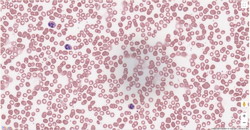
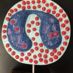
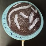
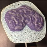
Use a human blood smear slide, focus under high power, then draw and label the formed elements of blood. It should look similar to what you see here. For online courses, use the University of Michigan Virtual Microscope, search for Human blood smear and complete the procedure as described for a regular microscope.

Procedure 3: Performing a Differential White Blood Cell Count
Obtain a human blood smear slide, focus under high power, count the number of white blood cells you see per field. Note and differentiate the type of WBC. Record your findings on the table provided. Calculate the % of each WBC present. For online courses, use the University of Michigan Virtual Microscope, search for Human blood smear and complete the procedure as described for a regular microscope.
| Cell type | Number of cells | % of WBC |
| Lymphocytes | ||
| Monocytes | ||
| Eosinophils | ||
| Basophils | ||
| Neutrophils |
Testing Simulated blood
Using a perform blood typing using a “Blood Typing Simulated Blood Kit”. Follow the instructions of the manufacturer.
For 100% online courses, please watch the blood typing simulation video.
Blood Vessels Lab
Introduction
The human circulatory system is a closed system composed of blood vessels, the heart and blood.
Recall what you learned about the directionality of blood in the unit of the heart, deoxygenated blood enters the right atrium via vena cava, and oxygenated blood enters the left atrium via pulmonary veins then the blood passes through the atrioventricular canals – to the right ventricle and to the left ventricle. When the ventricles of the heart contract, blood gets pushed in two different directions; from the right ventricle deoxygenated goes to the lungs – via the pulmonary trunk (artery) and return to the left atrium of the heart via pulmonary veins, then and from the left ventricle oxygenated blood is sent to the rest of the body via the aorta.
The blood will move into smaller arteries and finally into the capillaries (arterial end) in tissues, where exchange of materials will occur. The blood is then drain into the capillary bed (venous end) then into venules which will then empty into larger veins that will eventually reach the Vena Cava that enters the heart and completes the cycle.
The arteries are the ones that carry blood away from the heart and the veins will carry blood back to the heart. You usually see them colored in diagrams in red, blue and purple. The colors mean the following:
Red = blood vessels that carry oxygenated blood
Blue = blood vessels that carry deoxygenated blood
Purple = site for exchange where there is a mixture of oxygenated and deoxygenated blood or blood vessels that carry a mixture of both.
In this exercise we will examine and the gross and microscopic anatomy of selected blood vessels. We will identify mayor arteries and veins and learn which organs they are supplying or draining blood.
Learning Objectives
- Identify and label the components of blood vessels, arteries and veins.
- Identify selected arteries and veins.
- Describe the organs and regions supplied and drained by each artery and vein.
- Describe the blood flow patterns through portal systems.
- Trace the pathway of blood through pulmonary, cardiac, and systemic circuits.
- Describe the histological features of arteries, veins, and capillaries.
- Locate selected pulse points and identify locations for venipuncture.
Keywords
- macrovasculature
- microvasculature
- lumen
- tunica intima
- tunica media
- tunica adventitia
- elastic artery
- muscular artery
- continuous endothelium
- fenestrated endothelium
- discontinuous endothelium
- sinusoid
- artery
- arteriole
- vein
- venule
- capillary
- vasa vasorum
- portal system
Materials
- Anatomical models of the human circulatory system
- Anatomical Charts of human blood vessels (major arteries and veins)
- Diagrams of pulmonary, coronary and systemic circulation
- Anatomical model of a fetus with placenta
- Anatomical model of the brain – Arterial supply to the brain and sinuses
- Microscope slides:
- Cross section of artery and vein
- Heart muscle – intercalated disk preparation (capillaries)
- UM Virtual Microscope slide # 303, Artery & vein, Verhoeff stain, 40X.
Procedure 1 – Microscopic anatomy of blood vessels
Except for capillaries the walls of arteries and veins have several layers, these layers are called tunicas. There are three distinct tissue layers making up the walls of arteries and veins:
- Tunica interna or intima. It consists of a simple squamous epithelium (endothelium). It contains an adjacent basement membrane, the subendothelial connective tissue, and the internal elastic lamina. 2. Tunica media. Composed of smooth muscle tissue (innervated by the sympathetic nervous system) and elastic fibers, the elastic lamellae, where the elastic fibers allow the vessel to expand and recoil without damaging the walls of the arteries. 3. Tunica externa nor adventicia. The outermost layer of the blood, consists of dense irregular collagenous connective tissue with abundant collagen fibers, macrophages, mast cells, fibroblasts, nerves and vessels that supply large blood vessels.
- The image shown below was taken from the University of Michigan (UM) virtual Microscope, slide #303. Identify the artery and the vein.
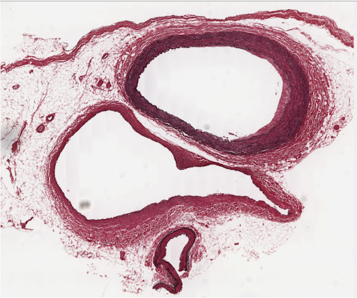
UM slide #303; Artery & vein, Verhoeff stain, 40X
- The image shown below was taken from the University of Michigan (UM) virtual Microscope, slide #303. Identify the artery and the vein.
Procedure 2 – Classification of Blood vessels
Blood vessels are divided into two broad categories:
- Macrovasculature – composed of blood vessels that can be seen with the naked eye.
- Microvasculature – composed of blood vessels that are smaller than 100 microns may only be seen with the aid of a microscope.
Blood vessels can be classified as: elastic arteries (conducting arteries), muscular arteries (distributing arteries), arterioles, capillaries (4 to 10 microns in diameter), venules, and veins (small, medium, and large), and portal systems. Veins of small and medium size are characterized by a thin media containing only a few layers of smooth muscle cells and a large tunica adventitia, considerably thicker than the tunica media. The vasa vasorum, is a network of small vessels that supplies the cells of larger blood vessels, present in their adventitia layer.
Types of Capillaries
Continuous – Capillaries with a continuous endothelium are less permeable and are present in muscles, lung, connective tissue, and skin.
Fenestrated – Capillaries with a fenestrated endothelium have gaps between endothelial cells, but the basement membrane is still continuous. These are found in the renal glomeruli, endocrine glands, intestinal villi, and exocrine pancreas.
Discontinuous – Capillaries with a discontinuous endothelium have large gaps between cells and a discontinuous basement membrane. Such capillaries are called sinusoids and are found in the liver and in blood-forming and lymphoid organs. These capillaries play an important role in blood/interstitial fluid exchanges and explain the high degree of capillary permeability for water and water-soluble molecules, as well as plasma protein and hormones.
- Complete the following table
| Type of blood vessel | Name an example of each |
| Elastic arteries | |
| Muscular arteries – Torso | |
| Muscular arteries – Head | |
| Muscular arteries – Arm | |
| Veins – large (torso) | |
| Veins – Large (leg) | |
| Veins – Large (arm) | |
| Portal System (brain) | |
| Portal System (abdomen) | |
| Fenestrated capillaries | Location: |
| Discontinuous capillaries | Location: |
Procedure 3 – Blood Vessels Identification
Use the anatomical models available in the laboratory to identify following arteries. For 100% online courses use the diagrams provided.
Arteries of the Trunk
- Aorta
- Ascending aorta
- Aortic arch
- descending aorta
- Thoracic aorta
- Abdominal aorta
- Brachiocephalic trunk
- Subclavian artery
- Celiac trunk
- Common hepatic artery
- Splenic artery
- Left gastric artery
- Middle suprarenal arteries
- Renal arteries
- Superior mesenteric artery
- Gonadal arteries
- Inferior mesenteric artery
- Common iliac artery
- Internal iliac artery
- External iliac artery
Arteries of the Head and Neck
- Common carotid arteries
- Right common carotid artery
- Left common carotid artery
- External carotid artery
- Superficial temporal artery
- Internal carotid artery
- Vertebral artery
- Basilar artery
- Cerebral arterial circle
- Anterior communicating artery
- Posterior communicating arteries
Arteries of the Upper Limbs
- Axillary artery
- Brachial artery
- Radial artery
- Ulnar artery
Arteries of the Lower Limbs
- Femoral artery
- Popliteal artery
- Anterior tibial artery
- Posterior tibial artery
Veins of the Trunk
- Superior vena cava
- Inferior vena cava
- Azygos system
- Azygos vein
- Hemiazygos vein
- Accessory hemiazygos vein
- Renal vein
- Gonadal veins
- Suprarenal vein
- Splenic vein
- Gastric veins
- Superior mesenteric vein
- Inferior mesenteric vein
- Hepatic portal vein
- Hepatic veins
Veins of the Head and Neck
- Vertebral vein
- External jugular vein
- Internal jugular vein
- Dural sinuses
- Superior sagittal sinus
- Inferior sagittal sinus
- Straight sinus
- Transverse sinus
- Sigmoid sinus
- Cavernous sinus
Veins of the Upper Limbs
- Radial vein
- Ulnar vein
- Brachial vein
- Cephalic vein
- Median antebrachial vein
- Basilic vein
- Median cubital vein
- Axillary vein
- Subclavian vein
- Brachiocephalic vein
Veins of the Lower Limbs and Pelvis
- Anterior tibial vein
- Posterior tibial vein
- Popliteal vein
- Small saphenous vein
- Great saphenous vein
- Femoral vein
- Common iliac vein
- Internal iliac vein
- External iliac vein
Procedure 4 – Trace the path of blood
Trace the path of blood on each of the vascular circuits shown below. Use arrows.
- Systemic Circuit

- Pulmonary Circuit

- Coronary Circuit

Procedure 5 – Histology of the Blood Vessels

For 100% online courses, go to University of Michigan Virtual Microscope, search for slide #303 or use the image shown here.
For standard face to face or hybrid classes, obtain a prepared microscope slides of artery, vein and capillaries. Note – for capillaries use a heart slide (intercalated disk stain).
Use colored pencils to draw and label the following:
- Artery
- Tunica interna (endothelium)
- Tunica media: Smooth muscle & Elastic fibers
- Tunica externa
- Lumen
- Capillary
- Tunica interna
- Blood cell(s), if present
- Lumen
- Vein
- Tunica interna
- Tunica media
- Tunica externa
- Lumen
Procedure 6 – Pulse Palpation
Pulse palpation is the process of using your fingertips to feel pulse points. You will be able to assess the rate, rhythm, and regularity of the heartbeat. Common pulse points are radial, ulnar, brachial, carotid, temporal, femoral, popliteal, posterior tibial, and dorsalis pedis arteries.
- Wash your hands
- Lightly place your index finger and middle finger over the artery, increase the pressure slightly, but be careful not to press too hard, because you could cut off blood flow.
- Start with the radial, ulnar, brachial, carotid, temporal, femoral, popliteal, posterior tibial, and dorsalis pedis arteries. Do the exercise only to one side of the body at the time,
- The superficial veins of the arm, forearm, and hand provide multiple places that are relatively easy to access to blood. When performing a venipuncture, the most commonly used veins are the median cubital vein, the cephalic vein, or the basilic vein. When placing an intravenous catheter, generally one of the posterior veins of the hand is used.
- Locate the most common places to perform venipuncture in your lab partner. We will not do the actual venipuncture.
Pre-Lab Quiz
- List, in order, the specific segmental variations of blood vessels that a red blood cell would travel through on a continuous circuit beginning in the left ventricle and ending in the right atrium of the heart.

- The blood-brain barrier (BBB) separates the brain and spinal cord from the contents of the bloodstream. What type of endothelium would you expect to see in the capillaries of the BBB?

- As you move through the arterial system from large to small arteries, what change would you expect to see in the relative amounts of elastic tissue and smooth muscle cells?

- As you move from arteries to veins, what change would you expect to see in the relative sizes of the tunica intima, media, and adventitia?

- Which of the following is a common pulse point? (check all that apply)
-
- Subclavian artery.
- Femoral artery.
- Brachial artery.
- Dorsalis pedis artery.
- Temporal artery
Fill in the blanks
- _________ carry blood away from the heart to supply tissues
- _________ drain tissues and bring blood back to the heart
- One layer of _____________ epithelium forms the walls of capillaries
- The _________ is the largest artery of the body
- The ________________ is the longest vein of the human body
- The ______________ is a branch of the abdominal aorta that supplies most of the small intestine and the final first half of the large intestine
- In the developing fetus the umbilical __________ carry rich blood in nutrients and oxygen to the fetus.
- In the developing fetus there is a connection between the right and the left atria called ___________.
- And minutes after a baby is born that closes and becomes _______________.
.
Pre-Lab Quiz (Online Version)
Media Attributions
- Heart model © Amazon is licensed under a All Rights Reserved license
- Blank circle
- Blood smear © Maria Carles is licensed under a CC BY-SA (Attribution ShareAlike) license
- Blank circle © Maria Carles is licensed under a CC BY-SA (Attribution ShareAlike) license
- Private: Artery and the vein label © Michigan Medical School adapted by Maria Carles is licensed under a CC BY-SA (Attribution ShareAlike) license

