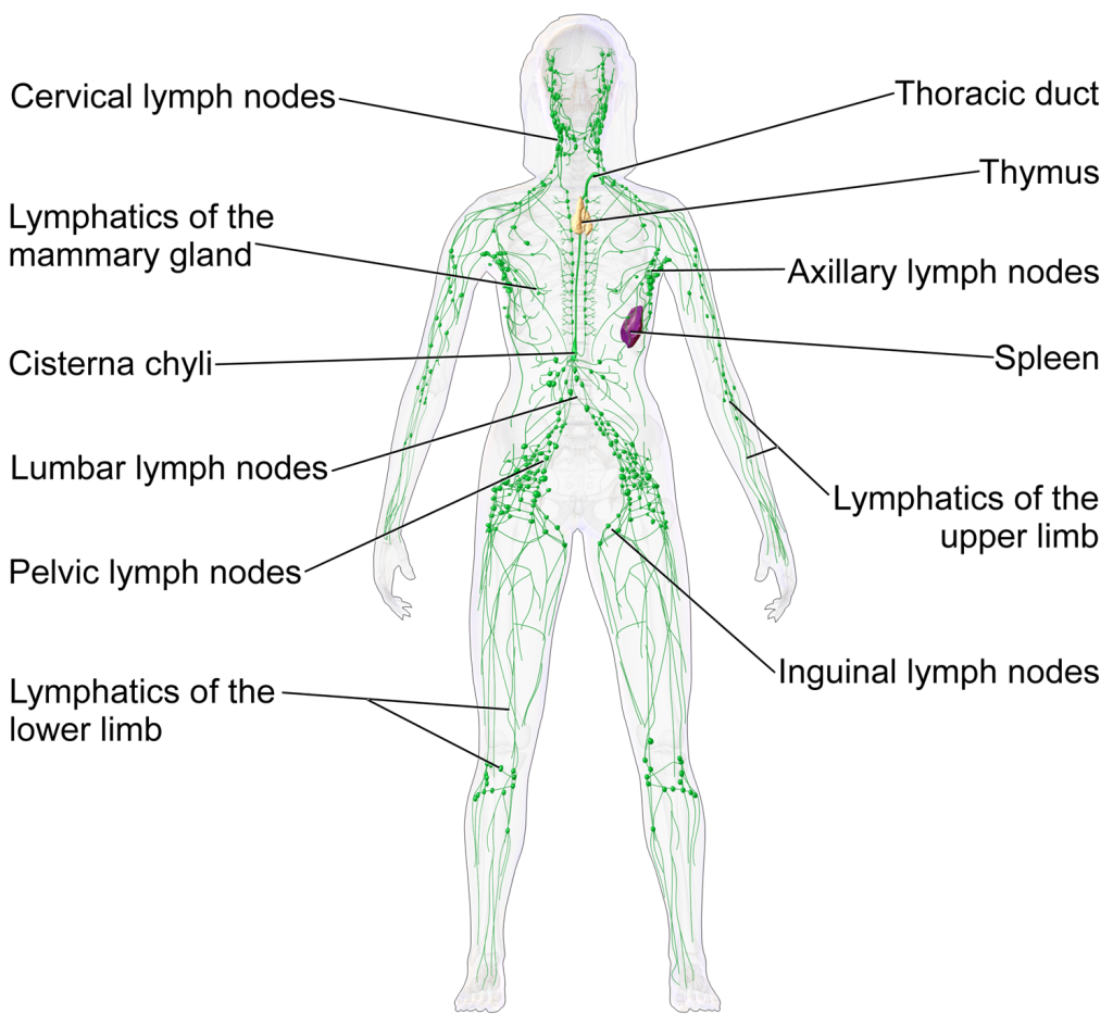
The Lymphatic and Immune System Anatomy
Human Lymphatic System

Introduction
The lymphatic system was first described in the 17th century independently by Olaus Rudbeck and Thomas Bartholin.
The lymphatic system is an organ system closely associated with the immune system, and complementary to the circulatory system. It consists of a large network of lymphatic vessels, lymph nodes, lymphoid organs, lymphoid tissues and lymph.
Lymph (Latin, lympha, it refers to the deity of fresh water, “Lympha”), is a clear fluid very similar in chemical composition to plasma, that is carried by the lymphatic vessels to the vena cava then into the heart for re-circulation. Lymph contains waste products and cellular debris, together with bacteria and proteins. The cells of the lymph are mostly lymphocytes.
The lymphoid organs include the lymph nodes (where the highest lymphocyte concentration is found), spleen, thymus, and tonsils. They are composed of lymphoid tissue and are the sites either of lymphocyte production or of lymphocyte activation. Lymphocytes are initially produced in the bone marrow and reach lymphoid organs via circulation. Lymphoid tissue may also be associated with the mucosa, mucosa-associated lymphoid tissue (MALT).
Fluid from circulating blood leaks into the tissues of the body by capillary action, carrying nutrients to the cells. The fluid bathes the tissues as interstitial fluid, collecting waste products, bacteria, and damaged cells, and then drains as lymph into the lymphatic capillaries and lymphatic vessels. These vessels carry the lymph throughout the body, passing through numerous lymph nodes which filter out unwanted materials such as bacteria and damaged cells. The lymph then passes into much larger lymph vessels known as lymphatic ducts; the right lymphatic duct drains the right side of the head and right arm and the larger left lymphatic duct, known as the thoracic duct, drains the left side of the body. The ducts empty into the subclavian veins, then to the superior vena cava to return to the fluid to the circulation. Lymph is moved through the system by muscle contractions.
Primary Lymphoid Organs
The primary lymphoid organs generate lymphocytes from immature progenitor cells. The thymus and the bone marrow constitute the primary lymphoid organs involved in the production and early clonal selection of lymphocyte tissues.
Bone Marrow
Bone marrow is responsible for both the creation of T cell precursors and the production and maturation of B cells, which are important lymphocyte types of the immune system. From the bone marrow, B cells immediately join the circulatory system and travel to secondary lymphoid organs in search of pathogens. T cells, on the other hand, travel from the bone marrow to the thymus, where they develop further and mature.
Thymus
The thymus increases in size from birth in response to postnatal antigen stimulation. It is most active during the neonatal and pre-adolescent periods. The thymus is located between the inferior neck and the superior thorax. At puberty, by the early teens, the thymus begins to atrophy and regress, with adipose tissue mostly replacing the thymic stroma. However, residual T cell lymphopoiesis continues throughout adult life, providing some immune response. The thymus is where the T lymphocytes mature and become immunocompetent. The loss or lack of the thymus results in severe immunodeficiency and subsequent high susceptibility to infection. In most species, the thymus consists of lobules divided by septa which are made up of epithelium which is often considered an epithelial organ. T cells mature from thymocytes, proliferate, and undergo a selection process in the thymic cortex before entering the medulla to interact with epithelial cells.
Secondary lymphoid organs
The secondary (or peripheral) lymphoid organs, which include lymph nodes and the spleen, maintain mature naive lymphocytes and initiate an adaptive immune response. The secondary lymphoid organs are the sites of lymphocyte activation by antigens. Activation leads to clonal expansion, and affinity maturation. Mature lymphocytes recirculate between the blood and the secondary lymphoid organs until they encounter their specific antigen.
Spleen
The main functions of the spleen are:
- to produce immune cells to fight antigens
- to remove particulate matter and aged blood cells, mainly red blood cells
- to produce blood cells during fetal life.
Lymph nodes
A lymph node is an organized collection of lymphoid tissue, through which the lymph passes on its way back to the blood. Lymph nodes are located at intervals along the lymphatic system. Several afferent lymph vessels bring in lymph, which percolates through the substance of the lymph node, and is then drained out by an efferent lymph vessel. Of the nearly 800 lymph nodes in the human body, about 300 are located in the head and neck. Many are grouped in clusters in different regions, as in the underarm and abdominal areas. Lymph node clusters are commonly found at the proximal ends of limbs (groin, armpits) and in the neck, where lymph is collected from regions of the body likely to sustain pathogen contamination from injuries. Lymph nodes are particularly numerous in the mediastinum in the chest, neck, pelvis, axilla, inguinal region, and in association with the blood vessels of the intestines.
The substance of a lymph node consists of lymphoid follicles in an outer portion called the cortex. The inner portion of the node is called the medulla, which is surrounded by the cortex on all sides except for a portion known as the hilum. The hilum presents as a depression on the surface of the lymph node, causing the otherwise spherical lymph node to be bean-shaped or ovoid. The different lymph vessel directly emerges from the lymph node at the hilum. The arteries and veins supplying the lymph node with blood enter and exit through the hilum. The region of the lymph node called the paracortex immediately surrounds the medulla. Unlike the cortex, which has mostly immature T cells, or thymocytes, the paracortex has a mixture of immature and mature T cells. Lymphocytes enter the lymph nodes through specialized high endothelial venules found in the paracortex.
A lymph follicle is a dense collection of lymphocytes, the number, size, and configuration of which change in accordance with the functional state of the lymph node. For example, the follicles expand significantly when encountering a foreign antigen. The selection of B cells, or B lymphocytes, occurs in the germinal center of the lymph nodes.
Secondary lymphoid tissue provides the environment for the foreign or altered native molecules (antigens) to interact with the lymphocytes. It is exemplified by the lymph nodes, and the lymphoid follicles in tonsils, Peyer’s patches, spleen, adenoids, skin, etc. that are associated with the mucosa-associated lymphoid tissue (MALT).
In the gastrointestinal wall, the appendix has mucosa resembling that of the colon, but here it is heavily infiltrated with lymphocytes.
Lymphatic vessels
The lymphatic vessels, also called lymph vessels, are thin-walled vessels that conduct lymph between different parts of the body. They include the tubular vessels of the lymph capillaries, and the larger collecting vessels–the right lymphatic duct and the thoracic duct (the left lymphatic duct). The lymph capillaries are mainly responsible for the absorption of interstitial fluid from the tissues, while lymph vessels propel the absorbed fluid forward into the larger collecting ducts, where it ultimately returns to the bloodstream via one of the subclavian veins.
The tissues of the lymphatic system are responsible for maintaining the balance of the body fluids. Its network of capillaries and collecting lymphatic vessels work to efficiently drain and transport extravasated fluid, along with proteins and antigens, back to the circulatory system. Numerous intraluminal valves in the vessels ensure a unidirectional flow of lymph without reflux. Two valve systems, a primary and a secondary valve system, are used to achieve this unidirectional flow. The capillaries are blind-ended, and the valves at the ends of capillaries use specialized junctions together with anchoring filaments to allow a unidirectional flow to the primary vessels. The collecting lymphatics, however, act to propel the lymph by the combined actions of the intraluminal valves and lymphatic muscle cells.
Lab Activity: Gross Anatomy of the Lymphatic System
Objectives
- List the main functions of the lymphatic system.
- List the components of the lymphatic system.
- Describe the relationship between the lymphatic system and the cardiovascular system.
- Describe the relationship between the lymphatic system and the immune system.
- Identify lymphatic organs and lymphatic nodules in the human body.
- Describe the microscopic structure of selected lymphatic structures. Describe the anatomy of an antibody molecule.
- Explain the basis of the ELISA test.
Purpose:
The goal of this lab is to examine the organization of the components of the lymphatic system. By the end of the lab, you should be able to describe and distinguish lymph, lymph nodes, tonsils, lymphatic tissue, thymus, and spleen.
Materials
- Torso
- Head and neck models
- Lymphatic system model
- Intestinal villus model (with lacteals)
Instructions
- 1. Obtain a model of the head and neck (midsagittal), identify the pharyngeal, lingual and palatine tonsils.
- Obtain a torso model, identify the thymus, spleen, ileum, and vermiform appendix.
- On a torso model, identify networks of lymphatic vessels in the axillary and inguinal regions, the right lymphatic duct, thoracic duct and cisternae chyli.
- Using a lymph node model, identify the afferent and efferent vessels, cortex and medulla.
- Using a lymphatic system model identify, cervical, axillary, inguinal, mediastinal, mesenteric lymph nodes, thoracic trunk, right lymphatic trunk, cisternae chili.
Microscopic Anatomy of the Lymphatic System Components
Materials
Microscope slides:
- Lymphatic vessels, l.s.
- Tonsils
- Thymus
- Lymph node
- Spleen
- Ilium (Peyer’s Patches)
Instructions
Lymphatic vessel, l.s.
- Obtain a slide of the lymphatic vessel, make sure it is a l.s. section.
- View the slide with the scanning objective to locate the sample, then change to low power. Identify the valves of the lymphatic vessels.
Tonsils
- Obtain a slide of the Tonsils.
- View the slide with the scanning objective to locate the sample, then change to low power. Identify the septa, germinal centers, cortex and medulla.
Thymus
- Obtain a slide of the thymus.
- View the slide with the scanning objective to locate the sample, then change to low power, then high power. Identify the capsule (connective tissue), and trabecula that subdivide each lobe of the thymus into lobules, cortex, medulla and germinal centers.
Lymph node
- Obtain a slide of the Lymph node.
- View the slide with the scanning objective to locate the sample, then change to low power and eventually to high power, 400X. Identify the capsule (connective tissue), cortex and medulla, germinal centers, afferent and efferent vessels, and septa.
Spleen
- Obtain a slide of the spleen.
- View the slide with the scanning objective to locate the sample, then change to low power, then high power. Identify the capsule and septa.
- Identify the two functional tissue components of the spleen; red pulp, and white pulp and the capsule.
Ilium patches
- Obtain a slide of Ilium.
- View the slide with the scanning objective to locate the sample, then change to low power. Identify the epithelium, the submucosa and the Peyer Patches
Anatomy of an Antibody
Draw and label an IgG molecule in the space provided.

Media Attributions
- Lymphatic system female © Wikipedia is licensed under a CC BY-SA (Attribution ShareAlike) license

