The Urinary System Anatomy
Introduction
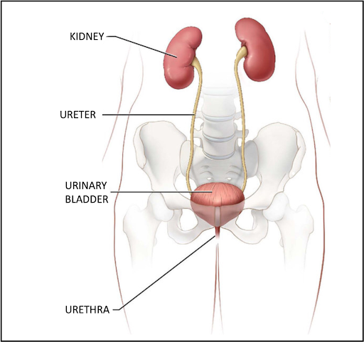
The urinary system consists of the two kidneys, two ureters, one urinary bladder, and one urethra.
The main functions of the urinary system and its components are to remove metabolic waste, regulate blood volume and composition (e.g., electrolytes, sodium, potassium, and calcium), regulate blood pressure, regulate pH of the blood, produce erythropoietin to signal production of red blood cells, and synthesize calcitriol (Vitamin D3).
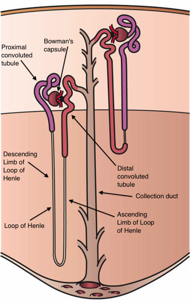
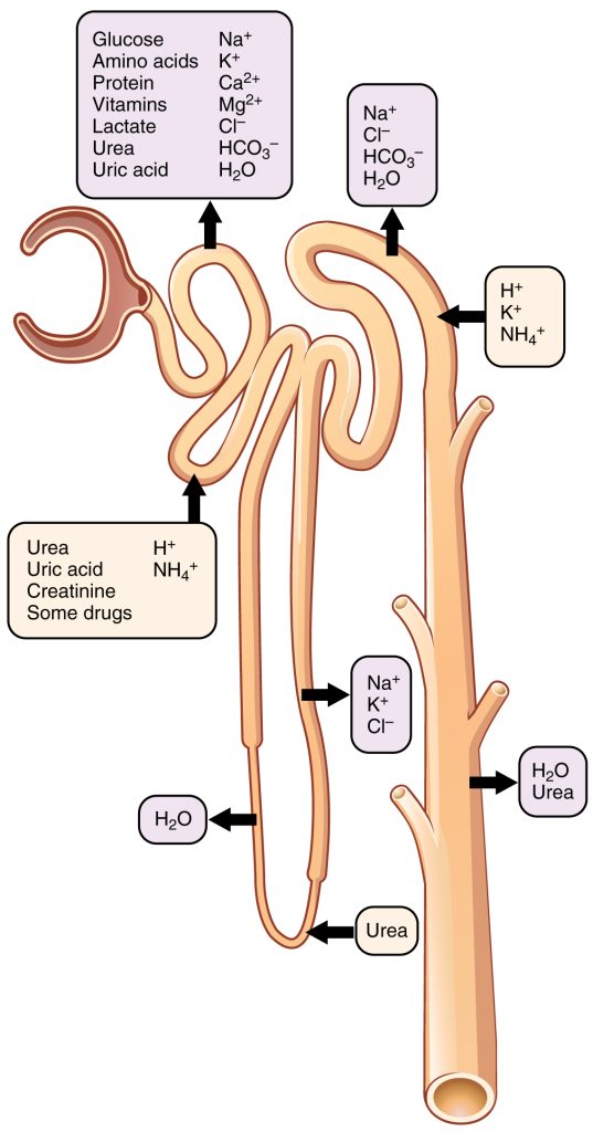
Each kidney consists of about one million functional units called nephrons, which are composed of a renal corpuscle (Bowman’s capsule and the glomerulus), a proximal convoluted tubule, a loop of Henle, and a distal convoluted tubule.
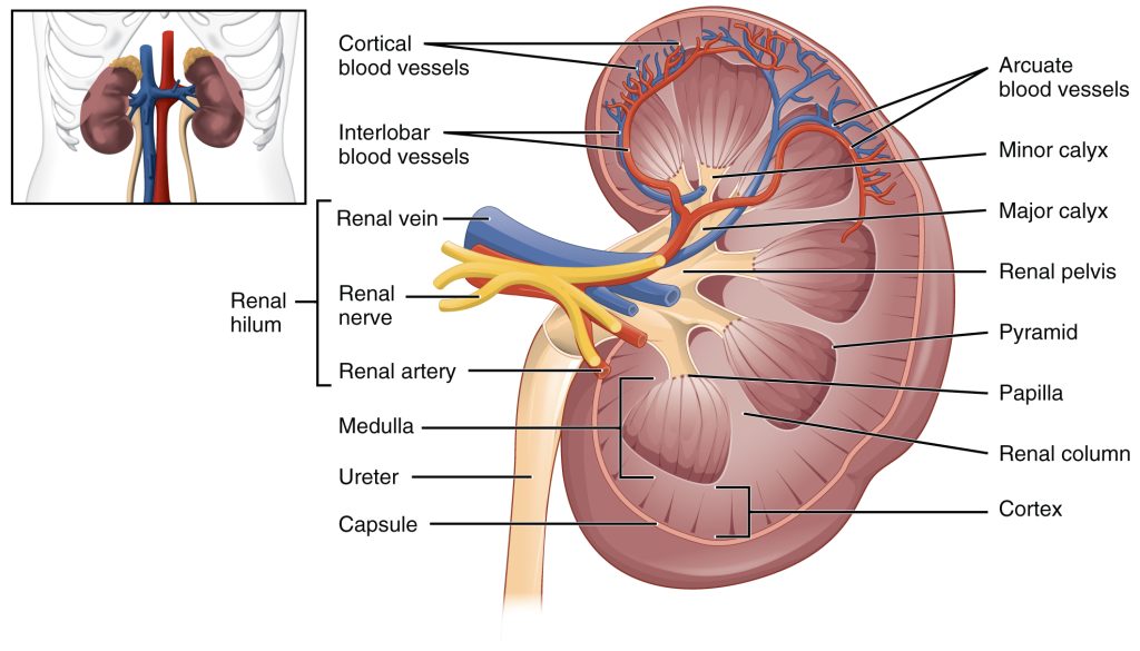
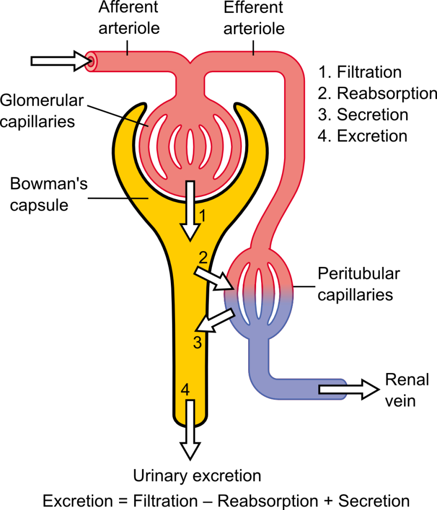
Kidneys are highly vascularized; blood enters the kidney via renal arteries (a branch from the abdominal aorta) and branches into the segmental, interlobar, arcuate, interlobular, and afferent arterioles. The afferent arterioles bring blood to the glomerulus (composed or fenestrated capillaries), where filtration occurs and blood exits the glomerulus via efferent arterioles. The efferent arterioles join the peritubular capillaries at the arterial end of the capillary bed, from there, blood continues to the venous end of the capillary bed that joins to the interlobular, arcuate, interlobar, segmental, and renal veins that bring blood to the inferior vena cava.
Processes of the Nephron
The nephrons are the functional units of the kidney and perform a series of processes. Filtration (1) starts at the glomerulus forming the filtrate will then pass to the proximal convoluted, loop of Henle, distal convoluted tubule, as the filtrate is passing through the tubules, reabsorption is taking place (2) as soon as the filtrate enters the proximal convoluted tubule, reabsorption begins and continues the length of the tubules including the collecting ducts. Most of the fluid is reabsorbed and the remaining secretion (3) is called urine. The urine is then transferred to the collecting ducts. Urine passes from the collecting duct to the minor calyces, then mayor calices, renal pelvis, and the ureter. The ureters (muscular tubes lined with transitional epithelium) propel urine towards the urinary bladder, the urine enter the posterior part of urinary bladder (two openings), where urine is stored until it is time to urinate. The urethra conveys the urine outside the body (4). The female and male urinary system are very similar in structure but differ in length, male urethra is about 4-5 times longer than the female urethra. An average of 800–2,000 milliliters (mL) of urine are normally produced every day in an adult; the final volume of urine varies according to fluid intake and kidney function.
In summary, the kidneys filtrate, reabsorb, secrete, and excrete waste products from our body.
Activity 1 – Introduction to the Urinary System Anatomy
Use the information shown in this YouTube video about the urinary system to complete this laboratory activity on the Urinary System. It may be helpful to pause the video to allow time to finish each part.
Activity 2 – Gross Anatomy of the Urinary System
Obtain a urinary system model from your instructor and identify the organs of the urinary system. You may use labeling tape and a black sharpie to label them. Please remember to remove the labels after you have shown it to your instructor.
Use this image shown below to label the organs of the urinary system. Enter the information on Table 1.
Table 1
| Number | Structure | Number | Structure |
| 1 | 8 | ||
| 2 | 9 | ||
| 3 | 10 | ||
| 4 | 11 | ||
| 5 | 12 | ||
| 6 | 13 | ||
| 7 | 14 |
For 100% online courses, skip using the urinary system model and use the drag and drop activity provided below to complete the labeling activities.
Kidneys are bean shaped structures located between the abdominal wall and the peritoneum; therefore, they are called retroperitoneal. They are attached to the abdominal wall
Use this image to label structures of the kidney. Enter the information on Table 2.
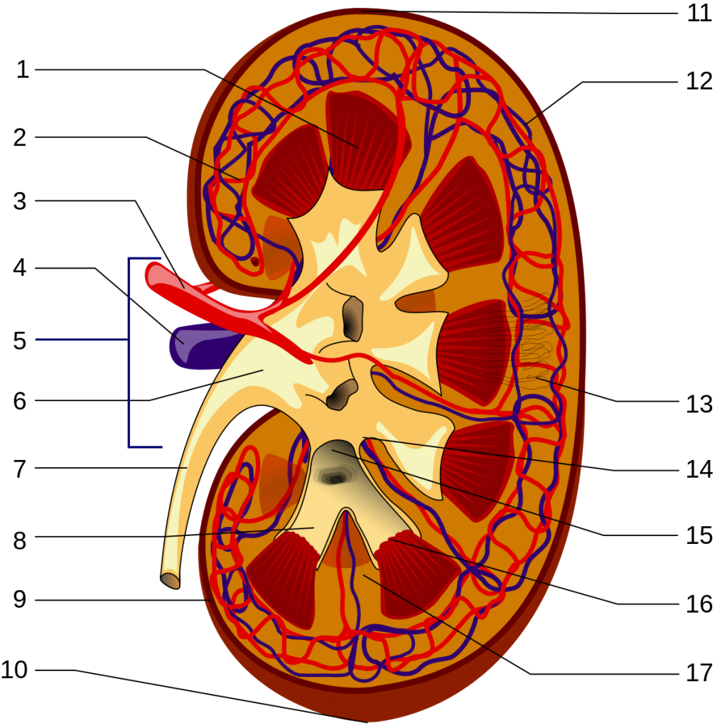
Table 2
| Number | Structure | Number | Structure |
| 1 | 10 | ||
| 2 | 11 | ||
| 3 | 12 | ||
| 4 | 13 | ||
| 5 | 14 | ||
| 6 | 15 | ||
| 7 | 16 | ||
| 8 | 17 | ||
| 9 |
For 100% online courses, skip using the structures of the kidney image and use the drag and drop activity provided below to complete the labeling activities.
Activity 3 – Microscopic Anatomy of the Urinary System
- For face to face or hybrid courses, obtain prepared slides of kidney, ureter and urinary bladder from our instructor. Draw and label them at 40X and a high magnification using the space provided (steps 5-7).
For 100% online courses, use the Virtual Microscope by following the instructions shown below: - Navigate to University of Michigan Virtual Microscope. – Full Slide List,
- In “Slide Category,” choose Urinary system.
- Chose slide #204 Kidney, human, H&E, 40X.
- Using scanning view locate the cortex and the medulla of the kidney.
- Increase magnification and locate the following:
-
- Glomerulus
- Proximal tubule
- Distal tubule
- Collecting duct
- Draw and label the kidney at 40X and a high magnification using the space provided.


- Chose slide #211 Ureter, adult human, H&E, 40X (transitional epithelium, smooth muscle).
- Using scanning view, locate the lumen and smooth muscle layer of the ureter.
- Increase magnification and locate the following:
-
- Lumen
- Mucosa (Transitional epithelium)
- Lamina propria
- Muscularis (smooth muscle; circular and longitudinal layers)
- Tunica adventitia (Fibrous connective tissue)
- Adipose tissue
- Draw and label the ureter at 40X and a high magnification using the space provided.


Chose slide #212 Bladder, human, H&E, 40X (transitional epithelium, smooth muscle).
- Navigate to https://histologyslides.med.umich.edu/Histology/Urinary%20System/212N_HISTO_40X.htm
- Using scanning view, locate the lumen and smooth muscle layer of the urinary bladder.
- Increase magnification and locate the following:
-
- Lumen
- Mucosa (Transitional epithelium)
- Lamina propria
- Detrusor muscle
- Draw and label the ureter at 40X and a high magnification using the space provided.


Answer the following questions:
- What is the composition of urine? (10 points)?

- Describe the four basic processes in the formation of urine (16 points).

Questions
- What is the composition of urine? (10 points)

- Describe the four basic processes in the formation of urine

Media Attributions
- Male and female bladder © Wikipedia is licensed under a CC BY-SA (Attribution ShareAlike) license
- Kidney nephron © Wikipedia is licensed under a CC BY-SA (Attribution ShareAlike) license
- 2610 the kidney © Wikipedia is licensed under a CC BY-SA (Attribution ShareAlike) license
- Physiology of nephron © Wikipedia is licensed under a CC BY-SA (Attribution ShareAlike) license
- Nephron secretion reabsorption © Wikipedia is licensed under a CC BY-SA (Attribution ShareAlike) license
- Urinary system © Wikipedia is licensed under a CC BY-SA (Attribution ShareAlike) license
- Kidney structures © Wikipedia
- Blank circle © Maria Carles is licensed under a CC BY-SA (Attribution ShareAlike) license

