The Reproductive System Anatomy
Introduction
The reproductive system in humans is complex. To our eyes the male and female reproductive systems very different but in reality, they are similar, they share the following characteristics:
- Both are derived from the same tissues during development.
- Contain gonads that produce gametes; two ovaries in females produce oocytes and two testes in males produce spermatozoids.
- Contain a system of ducts; to transport gametes to the site of fertilization; the epididymis, vas deferens and urethra in males and the oviducts in females.
- Contain accessory glands that secrete materials that aid in the transport of gametes to the site of fertilization. Seminal vesicles, prostate gland and bulbourethral gland in males and the greater vestibular glands in females.
- Their external genitalia contain similar structures, i.e. penis (male) and clitoris (female) and scrotal sac (males) and labial folds (female).
- Both sexes use the same hormones to regulate the reproductive process; Follicular stimulating hormone (FSH) and Luteinizing hormone (LH), both produced in the anterior pituitary. In males, interstitial cells from testes produce testosterone and in females, cells in the ovaries produce estrogen, both of these hormones are derived from cholesterol and have very similar chemical structure.
Even though these systems share many characteristics, they are fundamentally different and have different functions but they complement each other and work in close association for us to be able to generate the genetic diversity needed to perpetuate our species.
The male produces spermatozoids that contain half the number of chromosomes and the female produces oocytes that also contain half the number of chromosomes of human species. Through the process of fertilization, the genetic material will be combined as the oocyte and the sperm join to form a zygote. The zygote will then contain a complete set of chromosomes, half coming from the mother and the other half from the father. Once fertilization has occurred inside the female reproductive system, the zygote will implant in the innermost lining of the uterus, the endometrium where it will develop through numerous cell divisions into an embryo and then a fetus. The first stage of human development occurs around 6 days after fertilization and it is referred as pre-embryonic development (from zygote, 2-cell stage, 4-cells stage, morula, blastocyst) then it will continuous to develop into a fetus until the birth of a newborn.
Objectives
- Identify structures of the male reproductive system
- Identify structures of the female reproductive system
- Describe the histology of the testis (seminiferous tubules) and spermatozoa
- Describe the histology of the ovary
- Identify the stages of meiosis. Describe differences between mitosis and miosis
Laboratory Activities
Exercise 1: Define the following terms
Male Reproductive System
- Testes
- Seminiferous tubules
- Epididymis
- Ductus deferens
- Spermatic cord
- Seminal vesicle
- Prostate gland
- Corpus spongiosum
- Corpora cavernosa
- Testosterone
- Spermatogenesis
Female Reproductive System
- Ovaries
- Ovarian follicles
- Uterine tubes
- Uterus
- Cervix
- Vagina
- Vulva
- Mammary Glands
- Estrogen
- Oogenesis
Exercise 2 – Meiosis vs. Mitosis
- Draw and describe the stages of Meiosis
- List the differences between Mitosis and Meiosis
Exercise 3 – Identify reproductive organs using the anatomical models available in the laboratory
Use coronal and sagittal male pelvis models to identify the following structures:
Male Reproductive System
- Scrotum
- Testes
- Epididymis
- Seminiferous tubules
- Vas deferens
- Ejaculatory Tract
- Seminal Vesicle
- Bulbourethral gland
- Prostate
- Prostatic Urethra
- Membranous Urethra
- Penis
- Prepuce
- Glans Penis
- Spongy Urethra
Female Reproductive System
- Ovaries (2)
- Uterine tubes (2)
- Uterus
- Vagina
- External genital organs – VULVA
- Mammary glands
- Mons Pubis
Exercise 4 – Label and color the structures
Male Reproductive System
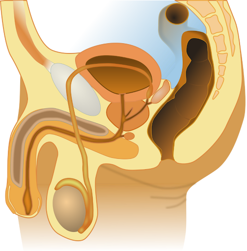
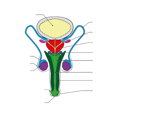
Label the male genital system using the following test bank
- Urinary bladder
- Vas deferens
- Seminal vesicle
- Prostate gland
- Bulbourethral gland or Cowper’s gland
- Bulb of penis (corpus spongiosum)
- Spongy Urethra
- Corpus Cavernosum
- Glans penis
- Urethral opening
- Foreskin or prepuce
- Epididymis
- Scrotum
- Testis
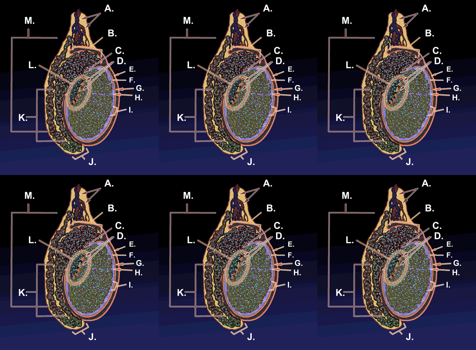
Label the structures of the Testis
| Letter | Structure |
| A | |
| B | |
| C | |
| D | |
| E | |
| F | |
| G | |
| H | |
| I | |
| J | |
| K | |
| L | |
| M |
Female Reproductive System
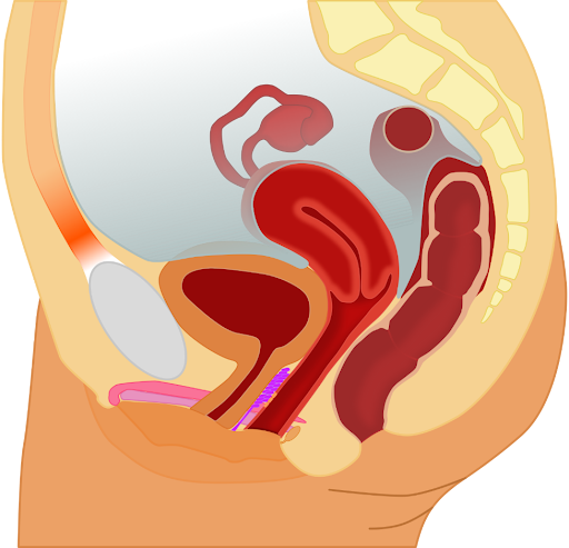
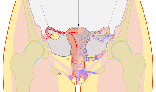
Label the structures using the word bank shown here:
- Vulva
- Labia majora
- Uterus
- Cervix
- Body Fundus
- Uterine cavity
- Endometrium
- Myometrium and Perimetrium
- Fallopian tube
- Isthmus; Ampulla
- Infundibulum
- Fimbria
- Ovary
- Broad ligament
- Round ligament
- Ovarian ligament
- Suspensory ligament of ovary
Exercise 5 – Microscopic Anatomy of Testes
Face-to-face Laboratories
Obtain a microscope slide of testes from your instructor and view using a standard light microscope. Identify, draw and label the following structures:
Seminiferous tubules
Interstitial cells (Leydig Cells)
Spermatogonia Sertoli cells
Spermatids
Lumen
Sperm in lumen of seminiferous tubules

Note: face to face labs should also take advantage of open educational resources, such as the virtual microscopes and review the images assigned to 100% onion labs.
100% online labs – University of Michigan Virtual Microscope – use the following link:
- Search for slide #70 Testis, human, H&E, 40X, you should see the following image at low magnification
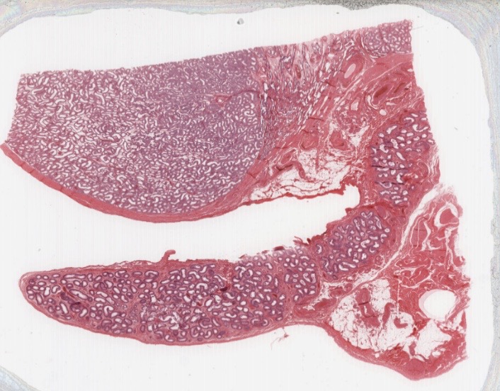
- Using the +/- controls, magnify the image to be able to identify and label the seminiferous tubules, Leydig cells (interstitial cells), lumen of the seminiferous tubules
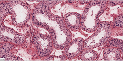
- Using the +/- controls, magnify even more the image to be able to identify and label the Leydig cells, Seminiferous tubules, Spermatogonia, Sertoli cells, spermatogonia, spermatids, sperm in lumen
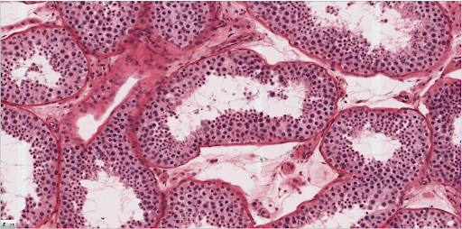
- Go to Slides for Junqueira’s Basic Histology
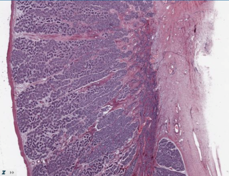
Slide No. 119, Testis, human H&E
Choose slide 119, testis, human H&E. Use the controls (+/-) to increase magnification. First scan the sample at low power and draw and label the structures of the testis:
- Capsule
- Lobules
- Convoluted seminiferous tubules

Increase magnification and draw and label the structures of the testis:
- Convoluted seminiferous tubules, x.s.
- Clusters of interstitial cells or Leydig Cells
- Spermatogonia
- Sertoli cells
- Spermatids
- Lumen
- Sperm (at lumen)

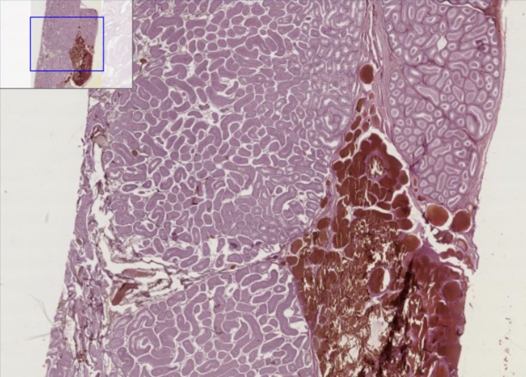
Choose slide 155, testis, dog. Use the controls (+/-) to increase magnification. First scan the area at low magnification, then increase magnification close to the maximum and draw and label the structures of the testis/seminiferous tubules:
- Seminiferous tubules (transverse section)
- Clusters of interstitial cells or Leydig Cells – between the seminiferous tubules
- Spermatogonia
- Sertoli cells
- Lumen
- Spermatids
- Sperm (at lumen)
Exercise 6 – Microscopic Anatomy of Ovaries
Face-to-face Laboratories
Obtain a microscope slide of ovary from your instructor and view using a standard light microscope. Identify, draw and label the following structures:
- Ovary
- Primordial Follicles
- Primary follicles
- Secondary follicles (sometimes these are hard to see)
- Tertiary Follicles
- Antrum
- Cumulative mass
- Oocyte
- Corpus luteum (if present)

Note: face to face labs should also take advantage of open educational resources, such as the virtual microscopes and review the images assigned to 100% onion labs.
100% online labs – University of Michigan Virtual Microscope – use the following link:
- Search for slide #239 Ovary, monkey, H&E, 40X, you should see the following image at low magnification
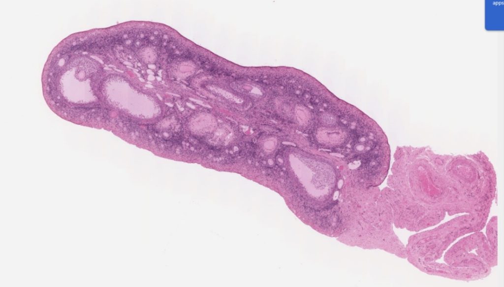
- Using the +/- controls, magnify the image to be able to identify and label the following structures: hilus, mesovarium, medulla, cortex, tunica albuginea, primordial follicles, ovum, granulosa
[follicular] cells, primary follicles, secondary follicles, antrum, mature [Graafian] follicles, theca interna, zona pellucida, cumulus oophorus, corona radiata, atresia, glassy membrane, corpus luteum, granulosa lutein cells, theca lutein cells, corpus albicans.
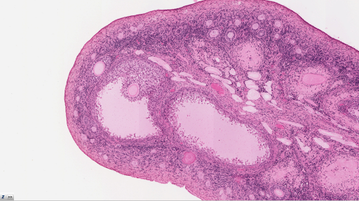
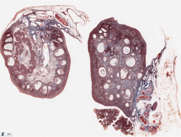
Choose slide 13, Ovary, Monkey, Masson Trichrome. Use the control (+/-) to magnify the image.
Draw, and label the following structures:
- Primordial follicles
- Primary follicles
- Secondary follicles
- Graafian follicles
- Oocyte
- Antrum
- Corpus luteum
- Corpus albicans
- Simple squamous epithelium Magnification ___________x
Media Attributions
- Label male reproductive system © Wikipedia is licensed under a CC BY-SA (Attribution ShareAlike) license
- Label male genital system front view © Maria Carles adapted by Wikipedia is licensed under a CC BY-SA (Attribution ShareAlike) license
- Testis © By KDS444 - Own work, CC BY-SA 3.0, https://commons.wikimedia.org/w/index.php?curid=35172098 is licensed under a CC BY-SA (Attribution ShareAlike) license
- Midsagittal section of female pelvis © Wikipedia is licensed under a CC BY-SA (Attribution ShareAlike) license
- Frontal section female reproductive organs © Wikipedia is licensed under a CC BY-SA (Attribution ShareAlike) license
- Blank circle © Maria Carles is licensed under a CC BY-SA (Attribution ShareAlike) license
- Testis, human, H&E, 40X © Maria Carles adapted by Michigan Medical School is licensed under a CC BY-NC-SA (Attribution NonCommercial ShareAlike) license
- Testis, human, Magnified 1 © Maria Carles adapted by Michigan Medical School is licensed under a CC BY-NC-SA (Attribution NonCommercial ShareAlike) license
- Testis, human, Magnified 2 © Maria Carles adapted by Michigan Medical School is licensed under a CC BY-NC-SA (Attribution NonCommercial ShareAlike) license
- No. 119, Testis, human H&E © Maria Carles adapted by Michigan Medical School is licensed under a CC BY-NC-SA (Attribution NonCommercial ShareAlike) license
- Testis, dog, H&E, Magnified © Maria Carles adapted by Michigan Medical School is licensed under a CC BY-NC-SA (Attribution NonCommercial ShareAlike) license
- Ovary, monkey, H&E, 40X © Maria Carles adapted by Michigan Medical School is licensed under a CC BY-NC-SA (Attribution NonCommercial ShareAlike) license
- Ovary, monkey magnified © Maria Carles adapted by Michigan Medical School is licensed under a CC BY-NC-SA (Attribution NonCommercial ShareAlike) license
- Ovary, Monkey, Masson Trichrome © Maria Carles adapted by Michigan Medical School is licensed under a CC BY-NC-SA (Attribution NonCommercial ShareAlike) license

