Chapter Four: Brain Development from Conception to Age 8
Biology of the Brain
In order to understand what a child’s brain can do, it is important to first understand how the brain is built. Throughout gestation and into the first months of life, the brain of the infant is growing. Perhaps you have heard that the human brain is not fully developed until age 25? This is true, but the majority of the cellular development takes place in the first three years of life so that the next 22 years can be spent refining how those cells are used. In this section, we will explore the smallest part of the brain – the cells – and how networks between cells develop and become the brain from a biological perspective.
There are six essential growth functions of your neural cells: cell proliferation, cell differentiation, cell migration, synaptogenesis, cell pruning, and myelination. In this section, you will learn the fundamentals of each of these processes, and throughout this textbook you will learn more about how these processes support children’s development in specific ages and stages. But first, let’s discuss what makes neural cells special, and how they work together to form the brain.
Brain Cells: Neurons
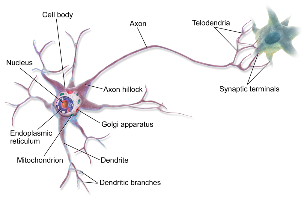
Like all living things, the brain is made of cells; brain cells are called neurons. Neurons are highly specialized cells that have three main parts: an axon, the cell body, and multiple dendrites. Electrical impulses run through these three structures in a specific pattern, causing chemical reactions which are our thoughts and feelings. These impulses also control our physical actions.

The cell body is just like any other cell in the human body: it has a nucleus that houses DNA and RNA, which is surrounded by cytoplasm that contains organelles. In this picture, do you recognize any of the cellular structures from biology class?
If it has been a while since you took Biology, or you just want a little more information, check out this Boundless Anatomy and Physiology course from Lumen!
From this image, you may also be noticing several differences between a brain cell and other cells in the body. The most obvious characteristics of neurons that set them apart from other cells are their dendrites and long axons.
In this illustration, the dendrites are the red branch-like structures extending from the main cell body. The dendrites are responsible for “touching” other cells – usually the axons of neighboring cells. You can see this depicted here where red branches are connecting to yellow branches, which come from a neighbor cell. Cells communicate with one another, passing along an electrical current that stimulates a chemical reaction. We perceive our thoughts and actions as instantaneous and smooth, but in reality our brains are constantly turning signals on and off in a wave like pattern!
At the site where the two cells meet, a gap called the synapse, electrical currents pass from one cell to another. This is simply the name for the junction site between an axon and a neighboring dendrite, but a lot of important works takes place at a synapse! Electrical impulses move along an axon and when it reaches the synaptic cleft, it needs to “jump” from one neuron to another. The electrical current is moved across through a chemical reaction that produces a neurotransmitter – literally a chemical that transmits impulses between neurons. This electrical impulse travels from the dendrite, through the cell body, and down the axon to the next neurons. This is what we refer to as brain cells “firing.”
Axons can be long relative to the size of an individual cell, allowing them to reach dendrites on neurons all around them. This can also make them fragile, so they need special protection (Konkel, 2018).
Imagine wires in your own home, constantly moving electricity around to where it needs to be. There are places where two wires meet and electricity is transmitted from one to the next, just like between two neurons. Also, like the wires in your house, the physical structure that contains the electricity needs to be insulated to keep the electrical current inside the system and moving to where it needs to go. Axons are insulated by means of a myelin sheath, which is a thin layer of lipids (fatty substance) that protects the axon and helps insulate the electrical impulse as it moves. Without a healthy myelin sheath, it is possible for impulses to be slowed down or even lost altogether.
Electrical currents run the length of the axon with the help of the myelin sheath (Jensen, 2019). This image shows two neurons: the one on the left does not have a myelin sheath, while the one on the right does. Axons without myelin can still conduct signals, but the impulses move much more slowly. Follow this link to see an animated version of these two neurons conducting an impulse.
Did you notice how much more slowly the impulse on the left was moving?
Imagine you and your friend are walking down the sidewalk; each square of pavement is a myelin node. If one of you walks while the other one jumps from square to square, who will get to the end of the block first? The person who is jumping! This is how myelin nodes are able to move electrical impulses so much more quickly.
They allow the current to “jump” along the axon, rather than moving in a slow, continuous wave.
Neural Networks
In the brain, single cells do have independent functions, but most neurons work in small clusters to accomplish a task or in larger clusters to control major functions of the body. For example, you have small but specialized areas of the brain that accept sensory information for each of the different parts of your body (see “temporal lobe” below) as well as an entire lobe that is devoted to visual information (see “occipital lobe” below). The size of an area and its density of neurons is related to how much work it needs to do. Generally speaking, we actively process and respond to significantly more visual information every day than we do to touch sensations.
Neural cells gather together to form brain structures automatically, following a genetic blueprint that is almost universal. This is called “experience independent” development (Berninger & Richards, 2002) because it doesn’t require any sort of environmental input or experience. However, large portions of the brain grow only through experience, either “expected” or “dependent.”
Expected experiences are those that the brain anticipates encountering – such as seeing the world for the first time- so it lays a foundation of neurons and then development continues based on what actually happens.
in the child’s life. “Experience dependent” neural networks are those which only develop if and when a child has an experience that leads to their creation.
For example, each person will develop a unique set of neural networks for their pets, their family members, their favorite ice cream sundae, and so on. While many of us might have similar networks, they will never be identical because not only do those networks consist of the memory of the specific thing (dog, mom, ice cream) they also consist of all of the emotions, language, and episodic memories that go with it.
Episodic memories are memories of things that have happened to us; they are narrative in nature (they are a story) and often are tied to strong visual, olfactory, and sensory memories.
As brain cells develop in utero, they become specialized for certain areas of the brain and follow genetically laid plans to move to the right areas and connect to other cells. You will read more about this later in this chapter, where we specifically talk about brain development in utero, but first it is important to understand what the areas of the brain actually are!
Media Attributions
- Multipolar Neuron diagram © BruceBlaus via. Wikimedia Commons is licensed under a CC0 (Creative Commons Zero) license

