
Chapter Four: Brain Development from Conception to Age 8
Brain Development During Gestation
Over the course of gestation, the brain grows rapidly and new neural cells grow at a rate of 50,000 to 100,000 per second between the 5th and 20th weeks. (Berninger & Richards, 2002; p79) This is where the six processes of neural development begin. At full-term birth, much of the neural foundation is complete and what comes next is to add on to the existing structure through experience. During the first three years of life, the brain will grow rapidly, adding millions of neuronal connections as it stores information from nearly every sensory, emotional, or cognitive experience.
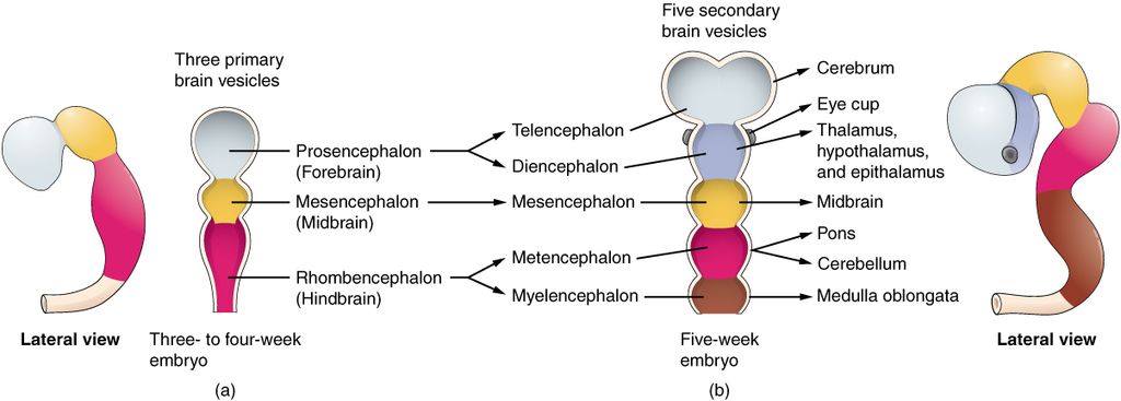
The brainstem, midbrain, and cerebral cortex will visibly develop during gestation. First, the brain stem develops, and it can be clearly distinguished from the other areas of the brain within the first month of gestation. At only four weeks into gestation, it is already possible to distinguish what will become the primary structures of the brain. This is due to rapid cell proliferation and differentiation. Proliferation is the generation of new neurons and their supporting cells, called glial cells. Differentiation refers to the shape and function of those cells, which become apparent as they grow.
As they grow, neurons move around in the brain so that they are physically located in the areas where they are functionally compatible. This process is called cell migration. Genetic information within the nucleus of each cell determines what its function will be and tells it where to move. Cells move by attaching to tracks created by glial cells (helper cells) that provide a track for the neurons’ journey. (Berninger & Richards, 2002; p 80) Typically, cells in the cerebral cortex migrate from the inner layers of the brain outward, building the brain from the inside/out – which should help to explain why the frontal lobe, somatosensory cortex, and even some areas related to language develop later than other areas of the brain.
The brainstem, the bottom-most section in the lateral view, is responsible for moving information between the body – where sensory information originates – and the brain – where it is processed and commands are given to the body. It connects the body to the three primary information processing areas of the brain: the cerebellum, the midbrain, and the cerebrum.
Brain development during gestation is influenced by a number of genetic and environmental factors. The overarching structure of the brain is independent of experience, being guided by genetic information from the beginning. Cell proliferation (the dividing of cells from a single cell into the trillions of cells that make up the human body) begins as soon as a fertilized egg embeds itself in the lining of the uterus, and cells continue to proliferate according to the instructions in each cell’s DNA. Of course, it is possible for DNA to give bad directions, and when this happens, the brain does not develop in the expected way. Environmental influences can damage DNA and cause irregularities in brain development. Perhaps one of the more well-known environmental causes of impaired brain development is from alcohol. Fetal Alcohol Spectrum Disorders are a family of diagnosable conditions that occur as a result of alcohol consumption during pregnancy. Although there are several physiological markers of FASDs, there are also many known cognitive side-effects due to the damage alcohol does to developing brain cells.
Alcohol consumption is not the only way a pregnant woman can influence the brain development of her child. General health and nutrition are important to ensure that the mother’s body is able to direct adequate resources to fueling cell proliferation and the building of fetal organs, including the brain. As we mentioned above, the long axons on the end of neurons are covered by a fatty sheath called myelin. The developing brain needs fat to build this sheath, so mothers need to supply this through their own diet.
Myelination of neural axons begins before birth, and at full gestation most of the spinal cord and brainstem are fully myelinated. However, myelination is not complete until adolescence! This means that although you may have the most neurons early in childhood, your brain works the quickest and most efficiently in your late teens and 20s. Throughout infancy, the midbrain and cerebellum are used regularly as infants learn to walk and talk; therefore, these areas receive the most myelination during the first two years of life. Yet the areas of the cerebral cortex, including the structures related to attention, language, and memory take up to ten years to be completely myelinated. Recall when you learned about the Emotional Domain of development, you read that the brain structures that underlie emotional regulation are not fully developed until later in life. This is because those areas are not fully myelinated, and myelination helps to strengthen their connections.
Myelination requires a healthy diet with plenty of healthy fats. You may already know that infants need a full fat diet, but since myelination continues for nearly 18 years, nutrition is important at all stages of life. Now, let’s think about how this relates to infants and their brain development. Have you ever seen a baby explore the world? What are some of the strategies that they use?
Media Attributions
- Brain Vesicle diagram © Openstax via. Wikimedia Commons is licensed under a CC BY (Attribution) license
