
The Origin of Eukaryotes
Section Goals
By the end of this section, you will be able to do the following:
- Describe what scientists know about the origins of eukaryotes based on the last common ancestor
- Explain the endosymbiotic theory
Before we discuss the origins of eukaryotes, it is first important to understand that all extant eukaryotes are likely the descendants of a chimera-like organism that was a composite of a host cell and the cell(s) of an alpha-proteobacterium that “took up residence” inside it. This major theme in the origin of eukaryotes is known as endosymbiosis, one cell engulfing another such that the engulfed cell survives and both cells benefit. Over many generations, a symbiotic relationship can result in two organisms that depend on each other so completely that neither could survive on its own.
Imagine you swallowed a small bird and suddenly gained the ability to fly, or you ate a cobra and were able to spit poisonous venom! Well, throughout the history of life, and specifically during the evolution of complex eukaryotic cells, things like this happened all the time. In fact, similar endosymbiotic associations are not uncommon even in living eukaryotes!
In the video below, science educator Adam Jacobson explains endosymbiosis.
Endosymbiotic events likely contributed to the origin of the last common ancestor of today’s eukaryotes and to later diversification in certain lineages of eukaryotes. But before explaining endosymbiosis further, it is necessary to consider metabolism in prokaryotes.
Prokaryotic Metabolism
Many important metabolic processes arose in prokaryotes; however, some of these processes, such as nitrogen fixation, are never found in eukaryotes. The process of aerobic respiration is found in all major lineages of eukaryotes, and it is localized in the mitochondria. Aerobic respiration is also found in many lineages of prokaryotes, but it is not present in all of them, and a great deal of evidence suggests that such anaerobic prokaryotes never carried out aerobic respiration, nor did their ancestors.
While today’s atmosphere is about 20 percent molecular oxygen (O2), geological evidence shows that it originally lacked O2. Without oxygen, aerobic respiration would not be expected, and living things would have relied on anaerobic respiration or the process of fermentation instead. At some point between 3.2 and 3.5 billion years ago, some prokaryotes began using energy from sunlight to power anabolic processes that reduce carbon dioxide to form organic compounds. That is, they evolved the ability to photosynthesize. Hydrogen, derived from various sources, was “captured” using light-powered reactions to reduce fixed carbon dioxide in the Calvin cycle. The group of Gram-negative bacteria that gave rise to cyanobacteria used water as the hydrogen source and released O2 as a “waste” product about 2.2 billion years ago.
Eventually, the amount of photosynthetic oxygen built up in some environments to levels that posed a risk to living organisms since it can damage many organic compounds. Various metabolic processes evolved that protected organisms from oxygen, one of which, aerobic respiration, also generated high levels of ATP. It became widely present among prokaryotes, including in a free-living group we now call alpha-proteobacteria. Organisms that did not acquire aerobic respiration had to remain in oxygen-free environments. Originally, oxygen-rich environments were likely localized around places where cyanobacteria were abundant and active. Still, by about 2 billion years ago, geological evidence shows that oxygen was building up to higher concentrations in the atmosphere. Oxygen levels similar to today’s levels only arose within the last 700 million years.
Recall that the first fossils that we believe to be eukaryotes date to about 2 billion years old, so they evolved and diversified rapidly as oxygen levels were increasing. Also, recall that all extant eukaryotes descended from an ancestor with mitochondria. These organelles were first observed by light microscopists in the late 1800s, where they were worm-shaped structures that were moving around in the cell. Some early observers suggested that they might be bacteria living inside host cells, but these hypotheses remained unknown or rejected in most scientific communities.
Endosymbiotic Theory
As cell biology developed in the twentieth century, it became clear that mitochondria were the organelles responsible for producing ATP using aerobic respiration, in which oxygen was the final electron acceptor.
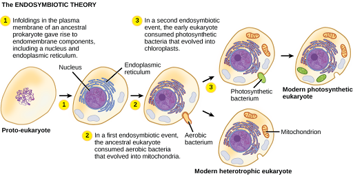
In the 1960s, American biologist Lynn Margulis of Boston University developed the endosymbiotic theory, which states that eukaryotes may have been a product of one cell engulfing another, one living within another, and coevolving over time until the separate cells were no longer recognizable as such and shared genetic control of a mutualistic metabolic pathway to produce ATP (Figure 3). In 1967, Margulis introduced new data to support her work on the theory and substantiated her findings through microbiological evidence. Although Margulis’s work initially was met with resistance, this basic component of this once-revolutionary hypothesis is now widely accepted, with work progressing on uncovering the steps involved in this evolutionary process and the key players involved.
While the metabolic organelles and genes responsible for many energy-harvesting processes appear to have had their origins in bacteria, our nuclear genes and the molecular machinery responsible for replication and expression appear to be more closely related to those found in the Archaea. Much remains to be clarified about how this relationship occurred; this continues to be an exciting field of Cooperation and Evolution Discovery in Biology. For instance, it is not known whether the endosymbiotic event that led to mitochondria occurred before or after the host cell had a nucleus. Such organisms would be among the extinct precursors of the last common ancestor of eukaryotes.
In this beautiful animation made from paper cutouts, Harvard Professor Rob Lue presents how mitochondria originated through endosymbiosis.
Mitochondria
One of the major features distinguishing prokaryotes from eukaryotes is the presence of mitochondria, or their reduced derivatives, in virtually all eukaryotic cells. Eukaryotic cells may contain anywhere from one to several thousand mitochondria, depending on the cell’s level of energy consumption, in humans being most abundant in the liver and skeletal muscles. Each mitochondrion measures 1 to 10 or greater micrometers in length and exists in the cell as an organelle. Although they may have originated as free-living aerobic organisms, mitochondria can no longer survive and reproduce outside the cell.
Mitochondria have several features that suggest their relationship to alpha-proteobacteria. Alpha-proteobacteria are a large group of bacteria that includes species symbiotic with plants, disease organisms that can infect humans via ticks, and many free-living species that use light for energy. Mitochondria have their own genomes, with a circular chromosome. Mitochondria also have special ribosomes and transfer RNAs that resemble these same components in prokaryotes. When mitochondrial genes are compared to those of other organisms, they appear to be of alpha-proteobacterial origin. In some eukaryotic groups, genes encoding proteins needed for cellular respiration are found in the mitochondria, whereas in other groups, they are found in the nucleus. This placement has been interpreted as evidence that over evolutionary time, genes have been transferred from the endosymbiont chromosome to those of the host genome. This apparent “loss” of genes by the endosymbiont is probably one explanation for why mitochondria cannot live without a host.
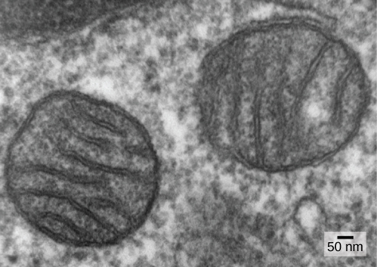
Another line of evidence supporting the idea that mitochondria were derived by endosymbiosis comes from the structure of the mitochondrion itself. Most mitochondria are shaped like alpha-proteobacteria and are surrounded by two membranes; the inner membrane is bacterial in nature, whereas the outer membrane is eukaryotic. This structure is exactly what one would expect if one membrane-bound organism was engulfed into a vacuole by another membrane-bound organism. The outer mitochondrial membrane was derived from the enclosing vesicle, while the inner membrane was derived from the plasma membrane of the endosymbiont. The mitochondrial inner membrane is extensive and involves substantial infoldings called cristae that resemble the textured outer surface of alpha-proteobacteria (Figure 1). The matrix and inner membrane are rich with the enzymes necessary for aerobic respiration.
The third line of evidence comes from the production of new mitochondria. Mitochondria divide independently by a process that resembles binary fission in prokaryotes. Mitochondria arise only from previous mitochondria; they are not formed from scratch (de novo) by the eukaryotic cell. Mitochondria may fuse together, and they may be moved around inside the cell by interactions with the cytoskeleton. They reproduce within their enclosing cell and are distributed with the cytoplasm when a cell divides or two cells fuse. Therefore, although these organelles are highly integrated into the eukaryotic cell, they still reproduce as if they were independent organisms within the cell. However, their reproduction is synchronized with the activity and division of the cell. These features all support the theory that mitochondria were once free-living prokaryotes.
Some living eukaryotes are anaerobic and cannot survive in the presence of too much oxygen. A few appear to lack organelles that could be recognized as mitochondria. In the 1970s and into the early 1990s, many biologists suggested that some of these eukaryotes were descended from ancestors whose lineages had diverged from the lineage of mitochondrion-containing eukaryotes before endosymbiosis occurred. Later findings suggest that reduced organelles are found in most if not all, anaerobic eukaryotes and that virtually all eukaryotes appear to carry some genes in their nuclei that are of mitochondrial origin.
In addition to the aerobic generation of ATP, mitochondria have several other metabolic functions. One of these functions is to generate clusters of iron and sulfur that are important cofactors of many enzymes. Such functions are often associated with the reduced mitochondrion-derived organelles of anaerobic eukaryotes. The protist Monocercomonoides, an inhabitant of vertebrate digestive tracts, appears to be an exception; it has no mitochondria, and its genome contains neither genes derived from mitochondria nor nuclear genes related to mitochondrial maintenance. However, it is related to other protists with reduced mitochondria and probably represents an end-point in mitochondrial reduction. Although most biologists accept that the last common ancestor of eukaryotes had mitochondria, it appears that the complex relationship between mitochondria and their host cell continues to evolve.
Plastids
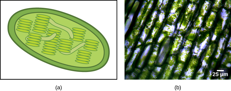
Some groups of eukaryotes are photosynthetic. Their cells contain, in addition to the standard eukaryotic organelles, another kind of organelle called a plastid. When such cells are carrying out photosynthesis, their plastids are rich in the pigment chlorophyll a and a range of other pigments, called accessory pigments, which are involved in harvesting energy from light. Photosynthetic plastids are called chloroplasts (Figure 2).
Like mitochondria, plastids have an endosymbiotic origin. This hypothesis was also proposed and championed with the first direct evidence by Lynn Margulis. We now know that plastids are derived from cyanobacteria that lived inside the cells of an ancestral, aerobic, heterotrophic eukaryote. This arrangement is called primary endosymbiosis, and two membranes surround plastids of primary origin. The common ancestor of the major lineage or “supergroup” Archaeplastida (containing red and green algae) took on a cyanobacterial endosymbiont (Figure 3). Almost all photosynthetic eukaryotes are descended from this event.
Cyanobacteria are a group of Gram-negative bacteria with all the conventional structures of the group. However, unlike most prokaryotes, they have extensive, internal membrane-bound sacs called thylakoids. Chlorophyll is a component of these membranes, as are many of the proteins of the light reactions of photosynthesis.
Chloroplasts of primary endosymbiotic origin have thylakoids, a circular DNA chromosome, and ribosomes similar to those of cyanobacteria. As in mitochondria, each chloroplast is surrounded by two membranes. The outer membrane is thought to be derived from the enclosing vacuole of the host, and the inner membrane is thought to be derived from the plasma membrane of the cyanobacterial endosymbiont.
There is also, as with the case of mitochondria, strong evidence that many of the genes of the endosymbiont were transferred to the nucleus. Plastids, like mitochondria, cannot live independently outside the host. In addition, like mitochondria, plastids are derived from the division of other plastids and are never built from scratch. Researchers have suggested that the endosymbiotic event that led to the Archaeplastida occurred 1 to 1.5 billion years ago, at least five hundred million years after the fossil record suggests that eukaryotes were present.
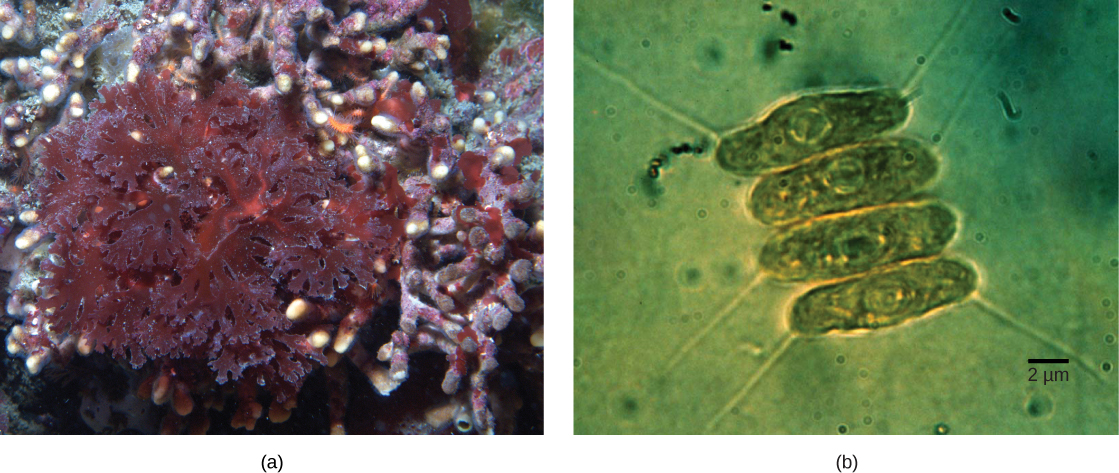
Not all plastids in eukaryotes are derived directly from primary endosymbiosis. Some of the major groups of algae became photosynthetic by secondary endosymbiosis, that is, by taking in either green algae or red algae (both from the group Archaeplastida) as endosymbionts (Figure 4). Numerous microscopic and genetic studies have supported this conclusion. Secondary plastids are surrounded by three or more membranes, and some secondary plastids even have clear remnants of the nucleus (nucleomorphs) of endosymbiotic algae. There are even cases where tertiary or higher-order endosymbiotic events are the best explanations for the features of some eukaryotic plastids.
Did I Get It?
CC Licensed Content, Shared Previously, Included in Introduction to the Organisms
- Biology 2e. Authors: Mary Ann Clark, Matthew Douglas and Jung Choi. Provided by: OpenStax CNX. Located at: Biology 2e. License: CC BY: Attribution 4.0.
- Biology for Majors II. Authors: Shelly Carter and Monisha Scott. Provided by: Lumen Learning. Located at: Biology for Majors II | Simple Book Production. License: CC BY: Attribution 4.0.
