
The Plant Body
Section Goals
By the end of this section, you will be able to do the following:
- Identify the different tissue types and organ systems in plants
- Describe the main function and basic structure of stems, roots, and leaves
Plants are as essential to human existence as land, water, and air. Without plants, our day-to-day lives would be impossible because, without oxygen from photosynthesis, aerobic life cannot be sustained. From providing food and shelter to serving as a source of medicines, oils, perfumes, and industrial products, plants provide humans with numerous valuable resources.
Watch Botany Without Borders, a video produced by the Botanical Society of America about the importance of plants.
When you think of plants, most of the organisms that come to mind are vascular plants. These plants have tissues that conduct food and water, and most of them have seeds. Seed plants are divided into gymnosperms and angiosperms. Gymnosperms include the needle-leaved conifers—spruce, fir, and pine—as well as less familiar plants, such as ginkgos and cycads. Their seeds are not enclosed by a fleshy fruit. Angiosperms, also called flowering plants, constitute the majority of seed plants. They include broadleaved trees (such as maple, oak, and elm), vegetables (such as potatoes, lettuce, and carrots), grasses, and plants known for the beauty of their flowers (roses, irises, and daffodils, for example).
While individual plant species are unique, all share a common structure: a plant body consisting of stems, roots, and leaves. They all transport water, minerals, and sugars produced through photosynthesis through the plant body in a similar manner. All plant species also respond to environmental factors, such as light, gravity, competition, temperature, and predation.
Like animals, plants contain cells with organelles in which specific metabolic activities take place. Unlike animals, however, plants use energy from sunlight to form sugars during photosynthesis. In addition, plant cells have cell walls, plastids, and a large central vacuole: structures that are not found in animal cells. Each of these cellular structures plays a specific role in plant structure and function.
Plant Tissues
Plants are multicellular eukaryotes with tissue systems made of various cell types that carry out specific functions. Plant tissue systems fall into one of two general types: meristematic tissue and permanent (or non-meristematic) tissue. Cells of the meristematic tissue are found in meristems, which are plant regions of continuous cell division and growth. Meristematic tissue cells are either undifferentiated or incompletely differentiated, and they continue to divide and contribute to the growth of the plant. In contrast, permanent tissue consists of plant cells that are no longer actively dividing.
Meristematic tissues consist of three types based on their location in the plant. Apical meristems contain meristematic tissue located at the tips of stems and roots, which enable a plant to extend in length. Lateral meristems facilitate growth in thickness or girth in a maturing plant. Intercalary meristems occur only in monocots, at the bases of leaf blades, and at nodes (the areas where leaves attach to a stem). This tissue enables the monocot leaf blade to increase in length from the leaf base; for example, it allows lawn grass leaves to elongate even after repeated mowing.
Meristems produce cells that quickly differentiate or specialize and become permanent tissue. Such cells take on specific roles and lose their ability to divide further. They differentiate into three main types: dermal, vascular, and ground tissue. Dermal tissue covers and protects the plant, and vascular tissue transports water, minerals, and sugars to different parts of the plant. Ground tissue serves as a site for photosynthesis, provides a supporting matrix for the vascular tissue, and helps to store water and sugars.
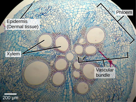
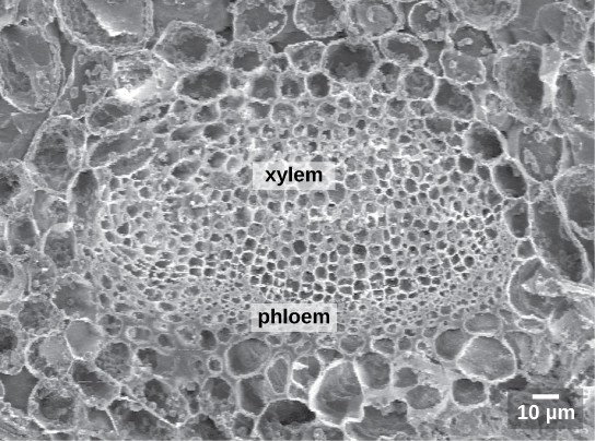
Secondary tissues are either simple (composed of similar cell types) or complex (composed of different cell types). Dermal tissue, for example, is a simple tissue that covers the outer surface of the plant and controls gas exchange. Vascular tissue is an example of a complex tissue and is made of two specialized conducting tissues: xylem and phloem. Xylem tissue transports water and nutrients from the roots to different parts of the plant. It includes three different cell types: vessel elements and tracheids (both of which conduct water), and xylem parenchyma. Phloem tissue, which transports organic compounds from the site of photosynthesis to other parts of the plant, consists of four different cell types: sieve cells (which conduct photosynthates), companion cells, phloem parenchyma, and phloem fibers. Unlike xylem-conducting cells, phloem-conducting cells are alive at maturity. The xylem and phloem always lie adjacent to each other (Figure 1). In stems, the xylem and the phloem form a structure called a vascular bundle.
Dermal Tissue
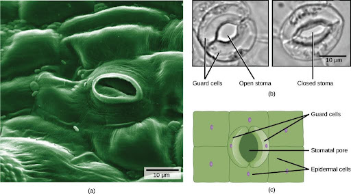
The dermal tissue of the stem consists primarily of epidermis, a single layer of cells covering and protecting the underlying tissue. Woody plants have a tough, waterproof outer layer of cork cells commonly known as bark, which further protects the plant from damage. Epidermal cells are the most numerous and least differentiated of the cells in the epidermis. The epidermis of a leaf also contains openings known as stomata, through which the exchange of gasses takes place (Figure 2). Two cells, known as guard cells, surround each leaf stoma, controlling its opening and closing and thus regulating the uptake of carbon dioxide and the release of oxygen and water vapor. Some leaves may have small hairs (trichomes) on the leaf surface. Trichomes help to deter herbivory by restricting insect movements, or by storing toxic or bad-tasting compounds; they can also reduce the rate of transpiration by blocking air flow across the leaf surface.
Vascular Tissue
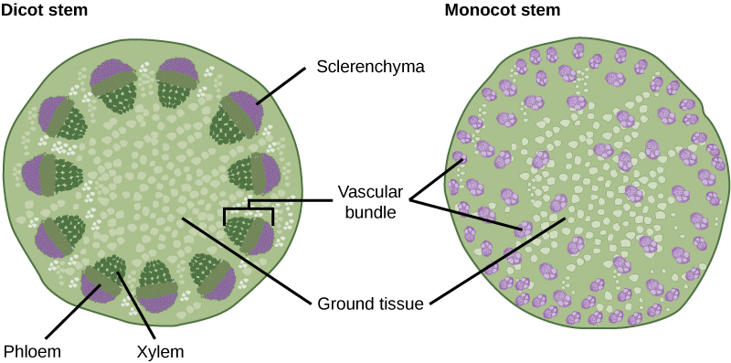
The xylem and phloem that make up the vascular tissue of the stem are arranged in distinct strands called vascular bundles, which run up and down the length of the stem. When the stem is viewed in cross-section, the vascular bundles of dicot stems are arranged in a ring. In plants with stems that live for more than one year, the individual bundles grow together and produce the characteristic growth rings. In monocot stems, the vascular bundles are randomly scattered throughout the ground tissue (Figure 3).
Xylem tissue has three types of cells: xylem parenchyma, tracheids, and vessel elements. The latter two types conduct water and are dead at maturity. Tracheids are xylem cells with thick secondary cell walls that are lignified. Water moves from one tracheid to another through regions on the side walls known as pits, where secondary walls are absent. Vessel elements are xylem cells with thinner walls; they are shorter than tracheids. Each vessel element is connected to the next by means of a perforation plate at the end walls of the element. Water moves through the perforation plates to travel up the plant. Phloem tissue is composed of sieve-tube cells, companion cells, phloem parenchyma, and phloem fibers. A series of sieve-tube cells (also called sieve-tube elements) are arranged end to end to make up a long sieve tube, which transports organic substances such as sugars and amino acids. The sugars flow from one sieve-tube cell to the next through perforated sieve plates, which are found at the end junctions between two cells. Although still alive at maturity, the nucleus and other cell components of the sieve-tube cells have disintegrated. Companion cells are found alongside the sieve-tube cells, providing them with metabolic support. The companion cells contain more ribosomes and mitochondria than the sieve-tube cells, which lack some cellular organelles.
Ground Tissue
Ground tissue is mostly made up of parenchyma cells but may also contain collenchyma and sclerenchyma cells that help support the stem. The ground tissue towards the interior of the vascular tissue in a stem or root is known as pith, while the layer of tissue between the vascular tissue and the epidermis is known as the cortex.
Plant Organ Systems
In plants, just as in animals, similar cells working together form a tissue. When different types of tissues work together to perform a unique function, they form an organ; organs working together form organ systems. Vascular plants have two distinct organ systems: a shoot system, and a root system. The shoot system consists of two portions: the vegetative (non-reproductive) parts of the plant, such as the leaves and the stems, and the reproductive parts of the plant, which include flowers and fruits. The shoot system generally grows above ground, where it absorbs the light needed for photosynthesis. The root system, which supports the plants and absorbs water and minerals, is usually underground. Figure 4 shows the organ systems of a typical plant.

Did I Get It?
Stems
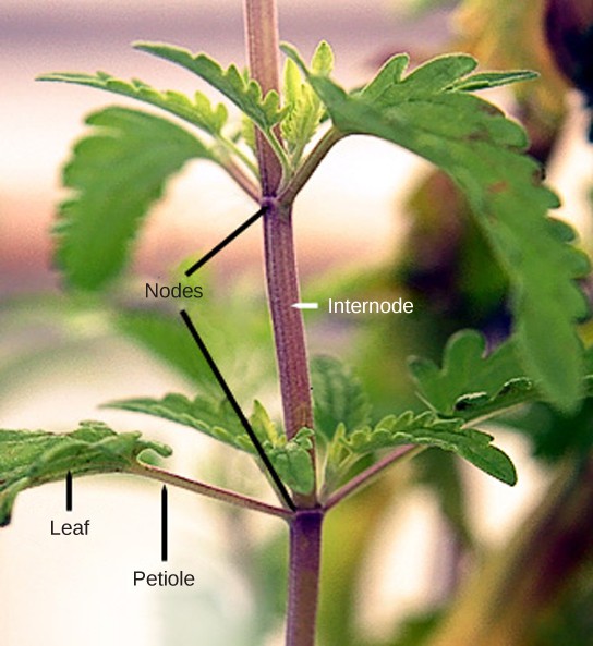
The stem of a plant bears the leaves, flowers, and fruits. Plant stems, whether above or below ground, are characterized by the presence of nodes and internodes. Nodes are points of attachment for leaves, aerial roots, and flowers. The stem region between two nodes is called an internode. The stalk that extends from the stem to the base of the leaf is the petiole (Figure 5).
Plant organs are made up of simple and complex tissues. The stem has three tissue systems: dermal, vascular, and ground tissue. Dermal tissue is the outer covering of the plant. It contains epidermal cells, stomata, guard cells, and trichomes. Vascular tissue is made up of xylem and phloem tissues and conducts water, minerals, and photosynthetic products. Ground tissue is responsible for photosynthesis and support.
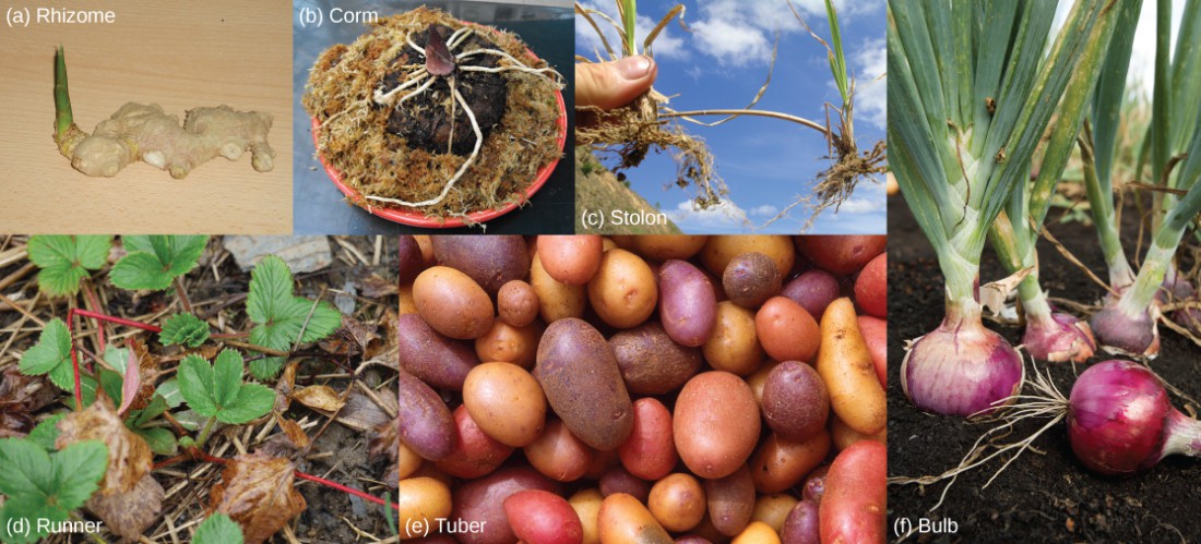
Primary growth occurs at the tips of roots and shoots, causing an increase in length. Woody plants may also exhibit secondary growth or increase in thickness. In woody plants, especially trees, annual rings may form as growth slows at the end of each season. Some plant species have modified stems that help to store food, propagate new plants, or discourage predators. Rhizomes, corms, stolons, runners, tubers, bulbs, tendrils, and thorns are examples of modified stems (Figure 6).
In the next video, watch botanist Wendy Hodgson of Desert Botanical Garden in Phoenix, Arizona, explain how agave plants were cultivated for food hundreds of years ago in the Arizona desert.
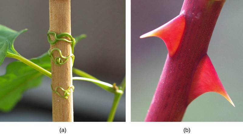
Some aerial modifications of stems are tendrils and thorns (Figure 7). Tendrils are slender, twining strands that enable a plant (like a vine or pumpkin) to seek support by climbing on other surfaces. Thorns are modified branches appearing as sharp outgrowths that protect the plant; common examples include roses, Osage orange and devil’s walking stick.
Roots
The roots of seed plants have three major functions: anchoring the plant to the soil, absorbing water and minerals and transporting them upwards, and storing the products of photosynthesis. Some roots are modified to absorb moisture and exchange gasses.
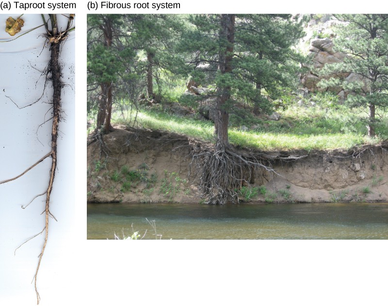
Taproots and fibrous roots are the two main types of root systems (Figure 8). In a taproot system, a main root grows vertically downward with a few lateral roots. Fibrous root systems arise at the base of the stem, where a cluster of roots forms a dense network that is shallower than a taproot.
The root has an outer layer of cells called the epidermis, which surrounds areas of ground tissue and vascular tissue. The epidermis provides protection and helps in absorption. Root hairs, which are extensions of root epidermal cells, increase the surface area of the root, greatly contributing to the absorption of water and minerals. Root vascular tissue conducts water, minerals, and sugars.
Inside the root, the ground tissue forms two regions: the cortex and the pith. Both regions include cells that store photosynthetic products. The cortex is between the epidermis and the vascular tissue, whereas the pith lies between the vascular tissue and the center of the root.
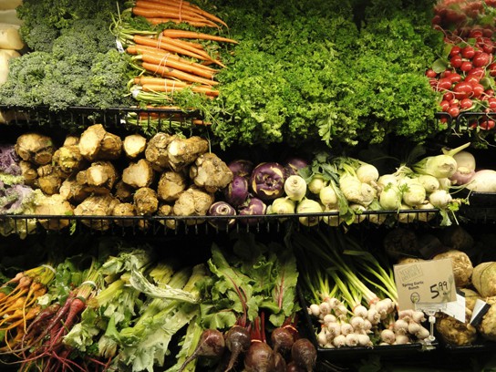
Root structures may be modified for specific purposes. For example, some roots are bulbous and store starch. Aerial roots and prop roots are two forms of aboveground roots that provide additional support to anchor the plant. Tap roots, such as carrots, turnips, and beets, are examples of roots that are modified for food storage (Figure 9).
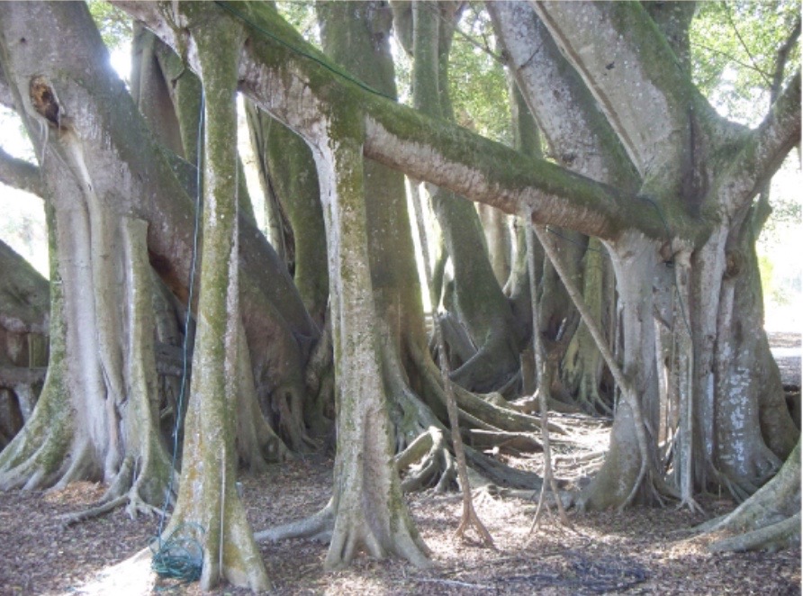
Epiphytic roots enable a plant to grow on another plant. The banyan tree (Ficus sp.) begins as an epiphyte, germinating in the branches of a host tree; aerial roots develop from the branches and eventually reach the ground, providing additional support (Figure 10).
Leaves
Leaves are the main site of photosynthesis. Most leaves are usually green, due to the presence of chlorophyll in the leaf cells. However, some leaves may have different colors, caused by other plant pigments that mask the green chlorophyll. A typical leaf consists of a lamina (the broad part of the leaf, also called the blade) and a petiole (the stalk that attaches the leaf to a stem).
The thickness, shape, and size of leaves are adapted to the environment. Each variation helps a plant species maximize its chances of survival in a particular habitat. Usually, the leaves of plants growing in tropical rainforests have larger surface areas than those of plants growing in deserts or very cold conditions, which are likely to have a smaller surface area to minimize water loss.
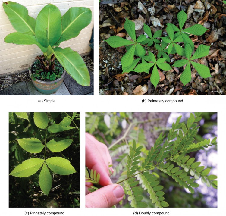
The arrangement of leaves on a stem, known as phyllotaxy, enables maximum exposure to sunlight. Each plant species has a characteristic leaf arrangement and form. The pattern of leaf arrangement may be alternate, opposite, or spiral, while leaf form may be simple or compound (Figure 11).
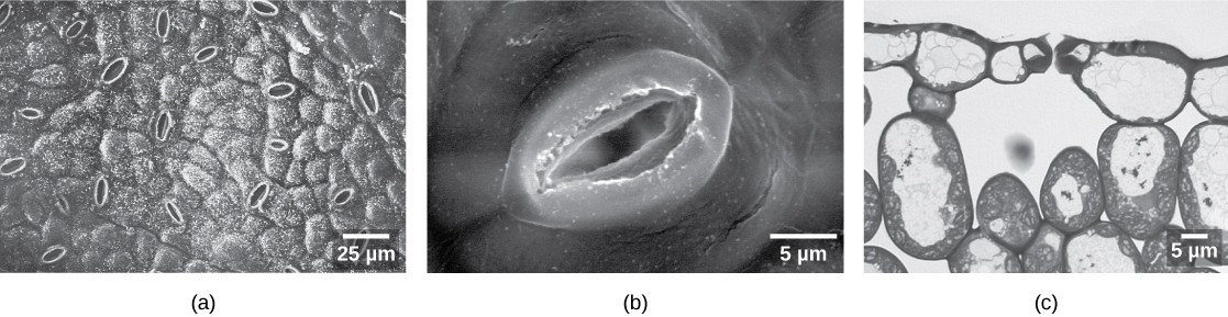
Leaf tissue consists of the epidermis, which forms the outermost cell layer, and mesophyll and vascular tissue, which make up the inner portion of the leaf. The epidermis helps in the regulation of gas exchange. It contains stomata (Figure 12), openings through which the exchange of gasses takes place. Two guard cells surround each stoma, regulating its opening and closing.
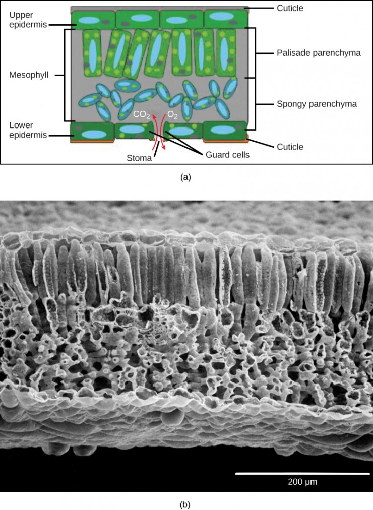
In the leaf drawing (Figure 13a), the central mesophyll is sandwiched between an upper and lower epidermis. The mesophyll has two layers: an upper palisade layer composed of tightly packed columnar cells, and a lower spongy layer, composed of loosely packed, irregularly shaped cells. Stomata on the leaf underside allow gas exchange. A waxy layer known as the cuticle covers the leaves of all plant species. The cuticle reduces the rate of water loss from the leaf surface. These leaf layers are clearly visible in the scanning electron micrograph (Figure 13b).
Like the stem, the leaf contains vascular bundles composed of xylem and phloem (Figure 14). The xylem consists of tracheids and vessels, which transport water and minerals to the leaves. The phloem transports the photosynthetic products from the leaf to the other parts of the plant. A single vascular bundle, no matter how large or small, always contains both xylem and phloem tissues.
In some plant species, leaf form is modified to form structures such as tendrils, spines, bud scales, and needles. Coniferous plant species that thrive in cold environments, like spruce, fir, and pine, have leaves that are reduced in size and needle-like in appearance. These needle-like leaves have sunken stomata and a smaller surface area, two attributes that aid in reducing water loss. In hot climates, plants such as cacti have leaves that are reduced to spines, which, in combination with their succulent stems, help to conserve water. Many aquatic plants have leaves with wide lamina that can float on the surface of the water and a thick waxy cuticle on the leaf surface that repels water.
Watch “The Pale Pitcher Plant” episode of the video series Plants Are Cool, Too, a Botanical Society of America video about a carnivorous plant species found in Louisiana.
Did I Get it?
CC Licensed Content, Shared Previously, Included in Plant Form and Physiology
- Biology 2e. Authors: Mary Ann Clark, Matthew Douglas, and Jung Choi. Provided by: OpenStax CNX. Located at: Biology 2e. License: CC BY: Attribution 4.0.
- Biology for Majors II. Authors: Shelly Carter and Monisha Scott. Provided by: Lumen Learning. Located at: Biology for Majors II | Simple Book Production. License: CC BY: Attribution 4.0.
