
10.2: Basic Emotions
Desire: The neural systems of reward-seeking
One of the most important affective neuronal systems relates to feelings of desire or the appetite for rewards. Researchers refer to these appetitive processes using terms such as “wanting” (Berridge & Kringelbach, 2008), “seeking” (Panksepp & Biven, 2012), or “behavioral activation sensitivity” (Gray, 1987). When the appetitive system is aroused, the organism shows enthusiasm, interest, and curiosity. These neural circuits motivate the animal to move through its environment in search of rewards such as appetizing foods, attractive sex partners, and other pleasurable stimuli. When the appetitive system is underaroused, the organism appears depressed and helpless.
Much evidence for the structures involved in this system comes from animal research using direct brain stimulation. When an electrode is implanted in the lateral hypothalamus or in cortical or mesencephalic regions to which the hypothalamus is connected, animals will press a lever to deliver electrical stimulation, suggesting that they find the stimulation pleasurable. Other regions in the desire system also include the amygdala, nucleus accumbens, and frontal cortex (Panksepp & Biven, 2012). The neurotransmitter dopamine, produced in the mesolimbic and mesocortical dopamine circuits, activates these regions. It creates a sense of excitement, meaningfulness, and anticipation. These structures are also sensitive to drugs such as cocaine and amphetamines, chemicals that have similar effects to dopamine (Panksepp & Biven, 2012).

Research in both humans and nonhuman animals shows that the left frontal cortex (compared to the right frontal cortex) is more active during appetitive emotions such as desire and interest. Early researchers noted that persons who suffered damage to the left frontal cortex developed depression, whereas those with damage to the right frontal cortex developed mania (Goldstein, 1939). The relationship between left frontal activation and approach-related emotions has been confirmed in healthy individuals using EEG and fMRI (Berkman & Lieberman, 2010). For example, increased left frontal activation occurs in 2- to 3-day-old infants when sucrose is placed on their tongues (Fox & Davidson, 1986), and in hungry adults as they view pictures of desirable desserts (Gable & Harmon-Jones, 2008). In addition, greater left frontal activity in appetitive situations has been found to relate to dopamine (Wacker et al., 2013).
“Liking”: The neural circuits of pleasure and enjoyment
Surprisingly, the amount of desire an individual feels toward a reward need not correspond to how much they like that reward. The neural structures responsible for enjoying rewards differ from those involved in desiring rewards. “Liking” (e.g., enjoyment of a sweet liquid) can be measured in babies and nonhuman animals by measuring licking speed, tongue protrusions, and happy facial expressions, whereas “wanting” (desire) is shown by the willingness to work hard to obtain a reward (Berridge & Kringelbach, 2008). Liking has been distinguished from wanting in research on topics such as drug abuse. For example, drug addicts often desire drugs even when they know that the ones available will not provide pleasure (Stewart et al., 1984).
Research on liking has focused on a small area within the nucleus accumbens and on the posterior half of the ventral pallidum. These brain regions are sensitive to opioids and endocannabinoids. Stimulation of other regions of the reward system increases wanting but does not increase liking and, in some cases, even decreases liking. The research on the distinction between desire and enjoyment contributes to the understanding of human addiction, particularly why individuals often continue to frantically pursue rewards such as cocaine, opiates, gambling, or sex, even when they no longer experience pleasure from obtaining these rewards due to habituation.
The experience of pleasure also involves the orbitofrontal cortex. Neurons in this region fire when monkeys taste or merely see pictures of desirable foods. In humans, this region is activated by pleasant stimuli, including money, pleasant smells, and attractive faces (Gottfried et al., 2002; O’Doherty et al., 2001; 2002; 2003).
Fear: The neural system of freezing and fleeing
Fear is an unpleasant emotion that motivates avoidance of potentially harmful situations. Slight stimulation of fear-related brain areas causes animals to freeze, whereas intense stimulation causes them to flee. The fear circuit extends from the central amygdala to the periaqueductal gray in the midbrain. These structures are sensitive to glutamate, corticotrophin-releasing factor, adreno-cortico-trophic hormone, and several different neuropeptides. Benzodiazepines and other tranquilizers inhibit activation in these areas (Panksepp & Biven, 2012).
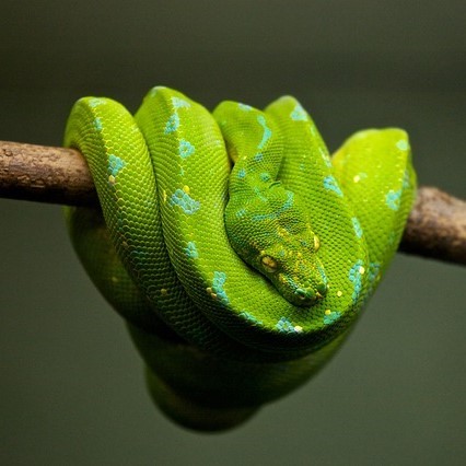
The role of the amygdala in fear responses has been extensively studied. Perhaps because fear is so important to survival, two pathways send signals to the amygdala from the sensory organs. When an individual sees a snake, for example, the sensory information travels from the eye to the thalamus and then to the visual cortex. The visual cortex sends the information on to the amygdala, provoking a fear response. However, the thalamus also quickly sends the information straight to the amygdala so that the organism can react before consciously perceiving the snake (LeDoux et al., 1990). The pathway from the thalamus to the amygdala is fast but less accurate than the slower pathway from the visual cortex. Damage to the amygdala or areas of the ventral hippocampus interferes with fear conditioning in both humans and nonhuman animals (LeDoux, 1996).
Rage: The circuits of anger and attack
Anger or rage is an arousing, unpleasant emotion that motivates organisms to approach and attack (Harmon-Jones et al., 2013). Anger can be evoked through goal frustration, physical pain, or physical restraint. In territorial animals, anger is provoked by a stranger entering the organism’s home territory (Blanchard & Blanchard, 2003). The neural networks for anger and fear are near one another but separate (Panksepp & Biven, 2012). They extend from the medial amygdala, through specific parts of the hypothalamus, and into the periaqueductal gray of the midbrain. The anger circuits are linked to the appetitive circuits, such that lack of an anticipated reward can provoke rage. In addition, when humans are angered, they show increased left frontal cortical activation, supporting the idea that anger is an approach-related emotion (Harmon-Jones et al., 2013). The neurotransmitters involved in rage are not yet well understood, but the neurotransmitter and neuromodulator Substance P (also involved in pain and stress) may play an important role (Panksepp & Biven, 2012). Other neurochemicals that may be involved in anger include testosterone (Peterson & Harmon-Jones, 2012) and arginine-vasopressin (Heinrichs et al., 2009). Several chemicals inhibit the rage system, including opioids and high doses of antipsychotics, such as chlorpromazine (Panksepp & Biven, 2012).
Love: The neural systems of care and attachment

For social animals such as humans, attachment to other members of the same species produces the positive emotions of attachment: love, warm feelings, and affection. The emotions that motivate nurturing behavior (e.g., maternal care) are distinguishable from those that motivate staying close to an attachment figure in order to receive care and protection (e.g., infant attachment). Important regions for maternal nurturing include the dorsal preoptic area (Numan & Insel, 2003) and the bed nucleus of the stria terminalis (Panksepp, 1998). These regions overlap with the areas involved in sexual desire and are sensitive to some of the same neurotransmitters, including oxytocin, arginine-vasopressin, and endogenous opioids (endorphins and enkephalins).
Grief: The neural networks of loneliness and panic
The neural networks involved in infant attachment are also sensitive to separation. These regions produce the painful emotions of grief, panic, and loneliness. When infant humans or other infant mammals are separated from their mothers, they produce distress vocalizations or crying. The attachment circuits are those that cause organisms to produce distress vocalizations when electrically stimulated.
The attachment system begins in the midbrain periaqueductal gray, very close to the area that produces physical pain responses, suggesting that it may have originated from the pain circuits (Panksepp, 1998). Separation distress can also be evoked by stimulating the dorsomedial thalamus, ventral septum, dorsal preoptic region, and areas in the bed nucleus of stria terminalis (near sexual and maternal circuits; Panksepp et al., 1988).
These regions are sensitive to endogenous opiates, oxytocin, and prolactin. All of these neurotransmitters prevent separation distress. Opiate drugs such as morphine and heroin, as well as nicotine, artificially produce feelings of pleasure and gratification similar to those normally produced during positive social interactions. This may explain why these drugs are addictive. Panic attacks appear to be an intense form of separation distress triggered by the attachment system, and panic can be relieved by opiates. Testosterone also reduces separation distress, perhaps by reducing attachment needs. Consistent with this, panic attacks are more common in women than in men.
Media Attributions
- Ice cream © Noba is licensed under a Public Domain license
- Snake © Noba is licensed under a Public Domain license
- In the mood for love © Flickr is licensed under a CC BY (Attribution) license
Part of the diencephalon. Regulates biological drives with pituitary gland.
The frontal cortex contains four main gyri.
A region of the basal forebrain located in front of the preoptic region.
A region of the frontal lobes of the brain above the eye sockets.
The gray matter in the midbrain near the cerebral aqueduct.
Almond-shaped neural structure that is primarily responsible for regulating emotional responses, especially fear.
A part of the diencephalon that works as a gateway for incoming and outgoing information.
The part of the brain that processes visual information that is located in the back of the brain.
A part of the anterior hypothalamus.
A band of fibers that runs along the top surface of the thalamus.
Originating from within an organism. For example, endogenous opioids such as endorphins are produced in the brain.
