
2.5: The Central Nervous System
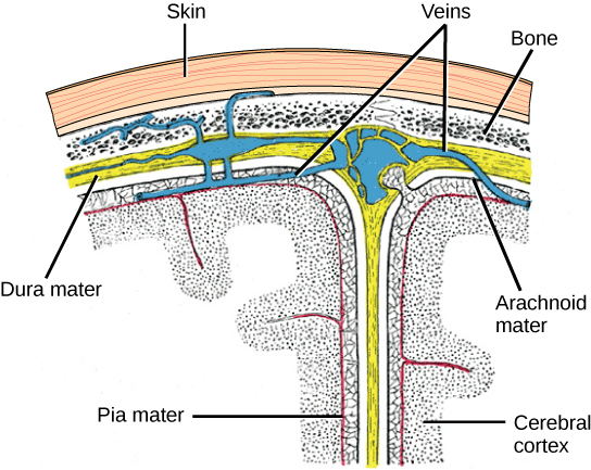
The central nervous system is made up of the brain and spinal cord. The brain and spinal cord are enclosed in three layers of protective coverings called meninges (from the Greek word for membrane) (Figure 8). The outermost layer is the dura mater (Latin for “hard mother”)—a thick layer that protects the brain and spinal cord and contains large blood vessels. The middle layer is the web-like arachnoid mater. The innermost layer is the pia mater (Latin for “soft mother”), which directly contacts and covers the brain and spinal cord like plastic wrap. The subarachnoid space between the arachnoid and pia maters is filled with cerebrospinal fluid (CSF), a fluid that helps cushion and protect the brain and spinal cord. We examine CSF more in the next section on The Brain.
The Spinal Cord
The spinal cord is the major bundle of nervous tissues that extends from the brainstem down the backbone to the lumbar region of the spine. The spinal cord transmits information from the skin, muscles, and internal organs to the brain, and vice versa. Information that travels from the bodily periphery toward the brain (or deeper centrally within the brain) is called an afferent signal. Information that travels away from the brain, such as a motor command to a muscle, is called an efferent signal.
In addition to sending information to and from the brain, the spinal cord controls some simple reflexive movements like removing your hand from a hot object and the knee reflex. These reflexive movements are very fast because the sensory signal is processed and the motor command is initiated directly in the spinal cord. Processing in the spinal cord avoids the time-consuming signal transmission to and from the brain, saving hundreds of milliseconds to protect our tissue from damage.
The spinal cord also houses central pattern generators that control some simple rhythmic movements such as walking. Experiments with cats have shown that even after severing the spinal cord (thereby cutting off motor commands from the brain), cats can still produce relatively normal walking on a treadmill (Duysens & Van de Crommert, 1998).
The spinal cord is protected by the bony vertebrae in the backbone and cushioned by cerebrospinal fluid. However spinal cord injuries still can occur and are very serious. In the United States, around 10,000 spinal cord injuries occur each year. Because the spinal cord is the information superhighway connecting the brain with the body, damage to the spinal cord can lead to paralysis. The extent of the paralysis depends on the location of the injury along the spinal cord and whether the spinal cord was completely severed. For example, if the spinal cord is damaged at the level of the neck, it can cause paralysis from the neck down, whereas damage further down the spinal column may limit paralysis to the legs. Spinal cord injuries are notoriously difficult to treat because spinal nerves do not regenerate, although ongoing research suggests that stem-cell transplants may be able to act as a bridge to reconnect severed nerves.
Text Attributions
This section contains material adapted from:
Clark, M.A., Douglas, M. & Choi, J. (2023). 35.3 The Central Nervous System. In Biology 2e. OpenStax. Access for free at https://openstax.org/books/biology-2e/pages/35-3-the-central-nervous-system License: CC BY 4.0 DEED.
Media Attributions
- The cerebral cortex © OpenStax is licensed under a CC BY-NC-SA (Attribution NonCommercial ShareAlike) license
The portion of the nervous system that includes the brain and spinal cord.
Signals that travel from the periphery toward the central nervous system and brain, or travel deeper centrally within the brain
Signals that travel away from the central nervous system or brain to the periphery, such as motor command
