
DNA Double-Helix Structure
Nucleotides thus come together to form long polymeric chains. In DNA, two chains pair antiparallel to one another via hydrogen bonds that link the bases in pairs. Antiparallel means that the two strands run in opposite directions, with the 5′ carbon end of one strand facing the 3′ carbon end of its matching strand, as shown in Figure 7.
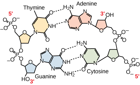
Only certain base pairings are allowed. Adenine can pair with thymine, and guanine can pair with cytosine, as Figure 6 shows. Adenine and thymine are thus said to be complementary, as are cytosine and guanine. Each base pair consists of one purine and one pyrimidine, which makes each base pair approximately the same size. We therefore also say that the DNA strands are complementary to each other.
If we know the sequence of one strand, we can always determine the sequence of the other. For example, if the sequence of one strand has the sequence 5’-AATTGGCC-3’, the complementary strand would have the sequence 3’-TTAACCGG-5’. We will see in the Replication and Transcription modules that this property of DNA makes it possible to both copy and use the genetic information stored in DNA.
A note on terminology: We are used to reading text left to right, from the top of a page to the bottom. But inside the cell, DNA is coiled around itself in three dimensions, and left/right and up/down do not have any meaning. Where would you start reading? So instead of thinking of DNA sequence from left-to-right, we think in terms of the 5’ and 3’ ends. The sequence 5’-AATTGGCC-3’ is the same as 3’-CCGGTTAA-5’, reading from the 5’ end through the bases to the 3’ end. For clarity, it is always best practice to label DNA sequence with the 5’ and 3’ ends. But by convention, if the 5’ and 3’ ends are not indicated, by convention it is assumed that the sequence is listed 5’ to 3’, left to right.
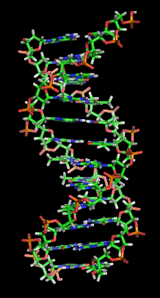
Under physiological conditions, the two paired chains coil around each other to form a double-helical molecule (Figure 8). The sugar and phosphate lie on the outside of the helix, forming the DNA’s backbone. The nitrogenous bases are stacked in the interior, like a pair of staircase steps. Hydrogen bonds bind the pairs to each other.
Media Attributions
- Antiparallel DNA strands © LibreTexts is licensed under a CC BY-SA (Attribution ShareAlike) license
- DNA double helix © Wikipedia is licensed under a CC BY-SA (Attribution ShareAlike) license
