Examples of tumor suppressor proteins
Most of the DNA repair proteins mentioned in the first half of this module are tumor suppressor proteins that act as caretakers. If DNA repair pathways fail, this results in a rapid accumulation of mutations. The rapid accumulation of mutations makes it more likely for a proto-oncogene to be mutated.
Two examples of gatekeeper tumor suppressors are Rb and p53. Rb was the first identified tumor suppressor, with mutations in Rb linked with the rare childhood cancer retinoblastoma. Rb mutations have since been found in other cancers as well[1]. Mutations in p53 are found in 50% of all human cancers. p53 plays a central role in multiple mechanisms of tumor suppression.
Rb
Retinoblastoma is a tumor of the retina. It is most diagnosed in children under the age of five. Historically, early stages of retinoblastoma were visible as white- or yellow-reflecting eyes in flash photography, shown on the left in Figure 15. While a healthy retina often produces a typical “red-eye” reflection in flash photography, the reflection of the flash against the tumor would produce a white or yellow reflection. (Note that with modern cell phone cameras the red-eye reduction features can produce yellow or white reflections too!) On the right in Figure 15 is the imaging of a patient’s retina, with the tumor visible as a lighter colored mass.

Figure 15. Image sources:
(left) http://visualsonline.cancer.gov/details.cfm?imageid=2418 , Public Domain; (right) Cieslik et al[2] https://www.researchgate.net/figure/Retinoblastoma-before-treatment-Visible-perforation-of-the-internal-limiting-membrane-in_fig1_371224804 CC BY 3.0
It most commonly occurs in children under five. The tumor can be seen in certain flash photographs as a white reflection in the eye as shown on the left. On the right is an image of the retina in a patient with retinoblastoma. The tumor is the light-colored mass within the red retinal tissue.
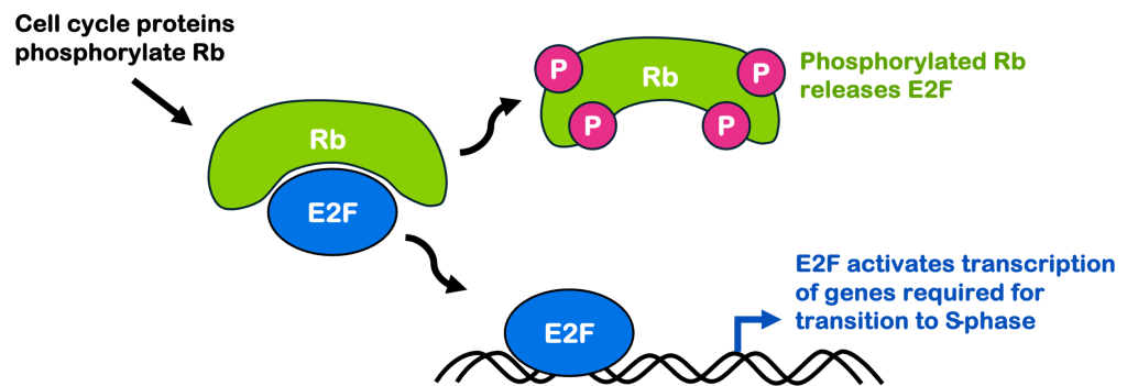
Retinoblastoma is caused by loss of function mutations in Rb. As shown in Figure 16, in healthy cells Rb blocks the transcription factor E2F. Cell cycle progression leads to the phosphorylation of Rb, which releases E2F. E2F transcribes genes required for the transition to S-phase of the cell cycle. A loss of function in Rb means E2F (and its downstream genes) are constantly active, leading to inappropriate cell cycling and proliferation. This is what leads to retinoblastoma tumors.
With modern medicine, most children in the United States with retinoblastoma are successfully treated through a combination of surgery and drug therapies.

p53
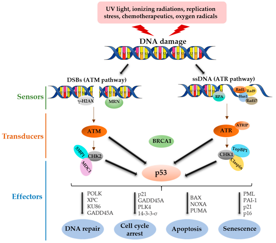
p53 has been called the “guardian of the genome”, with good reason. It can act as a sensor for DNA damage, relaying the signal to downstream targets and allowing the cell to respond in multiple ways. The cartoon in Figure 17, reprinted from a recent review article[3], is a bit complicated but gives a good overview of how many different ways p53 can act.
p53 is normally present at only low levels in the cell. DNA damage (shown at the top of the image) can be sensed by a variety of proteins in the cell. These are labeled “sensors” in this image. Those sensors relay a signal to transducers, which in turn stabilize p53 in the cell and allow it to accumulate at higher amounts. p53 is a transcription factor that, in turn, activates transcription of genes involved in DNA repair and cell cycle arrest.
p53 also can promote apoptosis and senescence. Apoptosis is a controlled form of cell death. A cell undertakes a series of molecular steps that will eventually trigger its own death. It might be counter-intuitive to think of cell death as a protective measure, but for a multicellular organism, the loss of one cell is not that big of a deal. It would be far worse for a damaged cell to become cancerous since that can potentially kill the whole organism. Senescence is when a cell permanently ceases cycling.
When a cell sustains low levels of DNA damage, p53 likely promotes cell cycle arrest and DNA repair, followed by re-entry into the cell cycle. If a cell sustains too much damage, though, p53 promotes apoptosis.[4]

Media Attributions
- Figure 15 DNA Repair combined © Cieslik et al is licensed under a CC BY-SA (Attribution ShareAlike) license
- Figure 16 DNA Repair © Amanda Simons is licensed under a CC BY-SA (Attribution ShareAlike) license
- Figure 17 DNA Repair © Gnanasundram et al. 2021.
- Engeland, K. Cell cycle regulation: p53-p21-RB signaling. Cell Death Differ. 29, 946–960 (2022). ↵
- Cieslik, K., Rogowska, A., Danowska, M. & Hautz, W. Efficacy of intravitreal injections of melphalan in the treatment of retinoblastoma vitreous seeding. Adv. Clin. Exp. Med. Off. Organ Wroclaw Med. Univ. 33, (2023). ↵
- Vadivel Gnanasundram, S., Bonczek, O., Wang, L., Chen, S. & Fahraeus, R. p53 mRNA Metabolism Links with the DNA Damage Response. Genes 12, 1446 (2021). ↵
- Chen, J. The Cell-Cycle Arrest and Apoptotic Functions of p53 in Tumor Initiation and Progression. Cold Spring Harb. Perspect. Med. 6, a026104 (2016). ↵

