
Geometry of the double helix
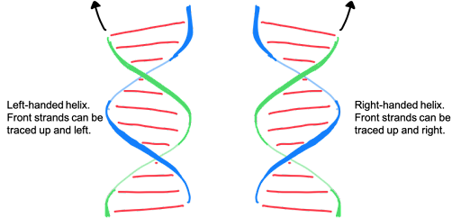
The X-ray diffraction images generated by Franklin, Wilkins, and others, allowed calculation of precise geometric arrangement of the atoms of the DNA molecule. In the double-helix, bases are paired together, stabilized by hydrogen bonds. The strands twist around each other counterclockwise, and they form a right-handed helix, as shown in figure 12 (right image).
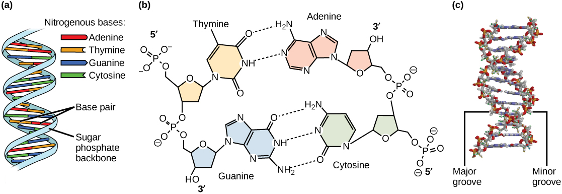
Remember that the two strands are anti-parallel in nature, with the 3′ end of one strand facing the 5′ end of the other strand. The sugar and phosphate of the nucleotides form the backbone of the structure, whereas the nitrogenous bases are stacked inside, like the rungs of a ladder. This is seen in Figure 13a, where the light blue ribbons of the backbone coil around the outside of the helix. One important feature to note is that adenine and thymine form two hydrogen bonds, while cytosine and guanine form three hydrogen bonds, as shown in Figure 13b. Each base pair is a flat structure, roughly trapezoidal in shape. The base pairs form the center of the helix, much like the steps of a spiral staircase, with the sugar-phosphate backbone forming the vertical railing around the outside.
Because each base pair is flat but shaped more like a trapezoid than a rectangle, the base pairs have different dimensions on each side relative to the phosphate groups. The twisting of the two strands around each other thus results in the formation of uniformly spaced major and minor grooves (Figure 13c). On the right face of the helix shown in Figure 13c, the minor groove is labeled. You can see that the red and yellow phosphate groups of the two strands are relatively close together, while on the left side, they are spaced farther apart (labeled as the major groove). This is also seen in Figure 13a, where the cartoon drawing shows the strands alternatingly farther apart and closer together.
The most common form of DNA found in a cell is called B-DNA. In B DNA, Each base pair is separated from the next base pair by a distance of 0.34 nm, and each turn of the helix measures 3.4 nm. Therefore, 10 base pairs are present per turn of the helix. The diameter of the DNA double-helix is 2 nm, and it is uniform throughout due to the consistent pairing of a larger purine with a smaller pyrimidine. These dimensions are also labeled in Figure 13.
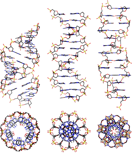
The structure we have been discussing, determined by Franklin, Watson, and Crick, is called B-DNA. The structure closely matches the structure of DNA found in the cell, which is an aqueous environment. Under other chemical conditions, DNA can assume slightly different conformations. A-DNA is a short, squat helix, with the bases slanted relative to the axis of the helix. It has a hollow core, as viewed down the center axis of the helix. It is shown on the left in Figure 14. A-DNA forms under dehydrating conditions, among others. DNA can even (very rarely) form a left-handed helix! The left-handed helix is called Z-DNA. Certain base sequences are more prone to forming Z-DNA. A-, B-, and Z-DNA are shown in Figure 14.

Media Attributions
- Left and right handed helix © Amanda Simons is licensed under a CC BY-NC-SA (Attribution NonCommercial ShareAlike) license
- A-, B-, and Z-DNA © Adapted from Biology LibreTexts, Working with Molecular Genetics (Hardison) is licensed under a CC BY-SA (Attribution ShareAlike) license
