
M phase: Mitosis
During mitosis, or M phase, the cell separates the duplicated copies of the genome and then separates the cytoplasm via cytokinesis. The mitotic cell cycle is how new somatic (nonreproductive) cells are generated in multicellular organisms. It is important during development and as cells are replenished due to age and injury in a mature organism.
The process of mitosis requires that each daughter cell have the same genome as the parent: exactly one copy of each chromosome. To make this happen, the cell keeps sister chromatids together after replication, connected via cohesion proteins. The chromatids then only separate during mitosis. Mitosis has four distinct stages: prophase, metaphase, anaphase, and telophase, with the physical division of the daughter cells, cytokinesis, beginning simultaneously with events of telophase. Some textbooks may count differently, including an intermediate prometaphase as a fifth stage, and some may count cytokinesis as a sixth. The stages of mitosis are shown in Figure 6.
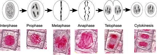
During interphase of the cell cycle, the nucleus is clearly visible within the cell. Individual chromosomes are not visible, and the nuclear membrane forms a barrier between DNA inside the nucleus and the cytosol. During the prophase of mitosis, the chromosomes condense, becoming more compact. At this stage, they begin to be visible under a microscope as individual chromosomes. The nuclear membrane dissolves, and there is no distinct separation from the cytosol.
In late prophase (sometimes distinguished as prometaphase), the chromosomes begin to migrate toward the center of the cell. In metaphase, the chromosomes align at the center (or equatorial plate) with one chromatid oriented toward each pole of the cell.
The movement and alignment of the chromosomes requires the action of the mitotic spindle. The mitotic spindle is made up of microtubules, long polymeric proteins which dynamically rearrange in mitosis to form the spindle. A cartoon of the spindle is shown in Figure 7. The microtubules (green) extend outward from microtubule organizing centers at the poles of each cell. Microtubules of the spindle attach to the chromosomes via kinetochore proteins (pink dots) that bind to chromosomes (blue) near the centromere. Spindle formation also requires the assembly of polar microtubules that extend outward from the poles but do not directly contact the chromosomes.

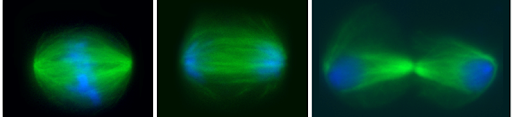
You can see the spindle via fluorescent microscopy in Figure 8. In Figure 8, microtubules are stained green and DNA is stained blue in sequential images that show metaphase, late anaphase, and cytokinesis. Note: The spindle is not seen in the cells in Figure 6, because the spindle proteins are not stained with the techniques used.
Once the chromosomes have assembled at the equatorial plate, the cohesins connecting the chromatids are dissolved. This allows the separation of chromatids, which are pulled to opposite poles of the cell by the mitotic spindle. This stage is called anaphase. In telophase, the separated chromatids (now unreplicated chromosomes) arrive at the poles of the cells, and nuclei reform.
Division of the cytoplasm is called cytokinesis, and it begins even as the separated chromatids are arriving at the poles. Because cytokinesis refers specifically to the splitting of the cytoplasm, it is considered distinct from telophase even though the two events happen simultaneously. Cytokinesis happens via different mechanisms dependent on cell type. Animal cells undergo cytokinesis as the cytosol is pinched off by a contractile ring of cytoskeletal proteins, as seen in the right panel of Figure 8. Plant cells separate by forming a new cell wall from vesicles that accumulate at the center of the cell. (This is seen in the telophase and cytokinesis cells shown in Figure 6.)
Accurate distribution of chromosomes to daughter cells requires the alignment of chromosomes at the equatorial plate, attachment to the mitotic spindle, and full separation of the chromatids. The M-phase checkpoint ensures that the chromosomes are properly attached to the spindle. If they are not properly attached, mitosis will pause.
