
Molecular genetics of epistasis
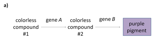
In most cases, the genetic interactions listed in Table 2 occur because the interacting genes are participating in a single biochemical pathway. Let’s look at a simple example of a pathway where two genes, A and B, act in sequence to produce a purple pigment (Figure 10).

So how does this all work for fur color in dogs? It all comes down to the molecular function of the genes. Let’s start with some basic biology.
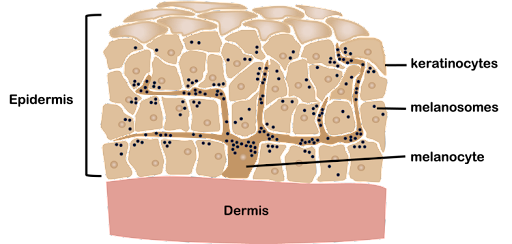
Pigmentation in mammals (including humans!) is primarily a result of two different forms of melanin: eumelanin is black or brown in color, while pheomelanin is reddish gold. Both eumelanin and pheomelanin are produced in specialized organelles called melanosomes within cells called melanocytes. Melanocytes are specialized cells with long arm-like projections that extend through the epidermis and contact many other cells (Figure 10). Melanosomes are transported to other cells in hair follicles and skin, giving hair (or fur, in the case of dogs) and skin its pigment. The ratio of eumelanin to pheomelanin, the amount of melanin in each melanosome, and the number of melanosomes produced by each melanocyte all influence overall pigmentation.
Each of the genes in Table 1 contributes to a different step of the pigmentation process. The B locus corresponds to a gene called TYRP1. This gene encodes an enzyme that catalyzes one of the first steps in synthesizing eumelanin from the amino acid tyrosine. In dogs, the B and b alleles produce slightly different forms of the enzyme. The enzyme variants produce slightly different eumelanin structures, which then appear either black or brown.
The melanosomes are transported through the branches of the melanosomes toward target cells in the epidermis, including hair follicles. A protein called melanophilin, encoded by the D locus, plays a role in this process. Different forms of the protein affect how many melanosomes are transported. Black or brown dogs with a dd genotype appear lighter in color because fewer melanosomes are transported. Which form of eumelanin (black or brown) is produced does not affect the action of melanophilin, which is why the B and D loci show no gene interaction in a dihybrid cross.
From the extensions of the melanocytes, melanosomes are targeted to other cells in the epidermis. The process is regulated by interactions between the melanocytes and the target cells. The E locus encodes a protein called MC1R. MC1R is a receptor protein found on the surface of melanocytes. In response to a signaling molecule called ASIP from hair follicles, MC1R triggers a switch between eumelanin and pheomelanin production in melanosomes destined for the fur. Variations in MC1R result in predominantly pheomelanin production (as can certain variations in the ASIP gene, also called the A locus)[1]. The reddish-color pheomelanin makes a dog appear yellow, cream, or red.
Do you have a yellow, cream, or red dog? Interestingly, you can tell your dog’s genotype at the B locus by looking at their nose! Although the e allele prevents eumelanin from being deposited in the fur, the black or brown pigment is still present in the skin of the nose. If your dog has a brown or pink nose, they have a bb genotype. If your dog has a black phenotype, they have at least one B allele, genotype B_[2].


As mentioned previously, dogs are a very powerful model organism because they are one of the most visibly variable species, but individual breeds may have very little genetic variation due to inbreeding. This makes it easier to identify genetic variants that correspond to traits, and it gives geneticists a “way in” to understanding human health as well. Many of the genes mentioned in this chapter perform similar functions in other mammals: including humans! Table 3 lists some of the genes controlling appearance in dogs, along with gene function and known phenotypic variations in humans.
| Gene name | Phenotypes in dogs | Phenotypes in humans | Gene function |
| KRT71
(Cu) |
Curly/smooth fur | One variant of KRT71 in humans affects hair texture. Variations in other types of keratin also affect hair texture in humans. | Encodes a protein from the keratin family that is a structural component of hair follicles. |
|---|---|---|---|
| TYRP1
(B) |
Black/brown | One variant of TYRP1 in humans is associated with blond hair in people of Melanesian ancestry. Other variants are associated with albinism[3]. | The encoded protein interacts with the enzyme that performs the first step in the synthesis of eumelanin. The b allele results in eumelanin that is brown in color, while the B allele results in eumelanin that is black. |
| MC1R
(E) |
Not yellow/yellow | Variations of MC1R in humans are associated with differences in skin and hair color[4]. | Encodes a protein on the surface of pigment-producing melanocytes. The e allele prevents the deposition of the black/brown eumelanin in the fur, leaving fur reddish/yellow due to the presence of pheomelanin. |
| MLPH
(D) |
Full color/faded or “dilute” pigment | A variation in MLPH is associated with Griscelli syndrome. People with Griscelli syndrome have hypopigmented skin and silvery hair[5]. | Encodes a protein called melanophilin, which plays a role in the transport of pigment-containing organelles within melanocytes. The recessive allele reduces the amount of eumelanin (black/brown pigment) that is deposited in fur. These alleles seem to affect eumelanin more than pheomelanin, so they have less of an effect on the color of a yellow/red dog with an ee genotype. |
| MITF
(S) |
No white/white spots or patches | Certain variants are associated with Waardenburg syndrome, which is characterized by areas of hypopigmentation and hearing loss. | Encodes a transcription factor that helps control the production of melanocytes (pigment-producing cells) |
| FGF5 | Short/long hair | A variation in FGF5 is associated with excessively long eyelashes[6]. | Encodes a growth factor that regulates the hair growth cycle in many mammals, including dogs and humans |
More than two genes
In most cases, the genetic interactions listed in Table 2 occur because the interacting genes are participating in a single biochemical pathway. But biochemical pathways often include far more than just two genes! The next sections look at how different forms of gene interaction influence phenotype when more than two genes are involved.
Media Attributions
- Purple pigment pathway © Amanda Simons is licensed under a CC BY-SA (Attribution ShareAlike) license
- Melanocytes © Amanda Simons is licensed under a CC BY-SA (Attribution ShareAlike) license
- Dog with brown nose © Joel Walsh is licensed under a CC BY-SA (Attribution ShareAlike) license
- Yellow dog black nose © Jennifer Sebalusky Williams is licensed under a CC BY-SA (Attribution ShareAlike) license
- Bannasch, D. L. et al. Dog colour patterns explained by modular promoters of ancient canid origin. Nat Ecol Evol 5, 1415–1423 (2021). ↵
- Schmutz, S. M., Berryere, T. G. & Goldfinch, A. D. TYRP1 and MC1R genotypes and their effects on coat color in dogs. Mamm Genome 13, 380–387 (2002). ↵
- TYRP1 gene: MedlinePlus Genetics. https://medlineplus.gov/genetics/gene/tyrp1/. ↵
- MC1R gene: MedlinePlus Genetics. https://medlineplus.gov/genetics/gene/mc1r/. ↵
- MLPH gene: MedlinePlus Genetics. https://medlineplus.gov/genetics/gene/mlph/. ↵
- Higgins, C. A. et al. FGF5 is a crucial regulator of hair length in humans. Proceedings of the National Academy of Sciences 111, 10648–10653 (2014). ↵
