
The cell cycle
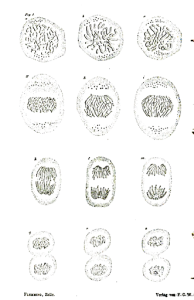
If living cells are observed under the microscope, they visibly change in morphology (size and shape) as they prepare for and then undergo cell division. One of the first biologists to observe this was Walther Fleming, who developed methods for staining cells that allowed him to see chromosomes. A cell actively undergoing division was easy to identify. It changed shape, pulling away from other nearby cells. The nucleus disappeared, but individual chromosomes became distinguishable from one another and moved through the cell in a distinctive pattern that was consistent from dividing cell to dividing cell. An example of hand-drawn images from his 1882 book, Zellsubstanz, Kern und Zelltheilung (Cell Substance, Nucleus, and Cell Division) is shown in Figure 1.
Fleming observed that the sequence of events drawn in Figure 1 all happened within a one-hour period, ending with the separation of two daughter cells. This cycle would repeat every 24 hours, but there was not much visible change in the intervening 23 hours. These two distinct phases of the cell became known as mitosis and interphase[1].
The most rapidly dividing eukaryotic cells take around 24 hours to divide, but in other cell types division might only occur every few days (or even weeks or months or not at all!). In those cells, interphase is lengthened, but the process of mitosis is consistently around 1-2 hours.
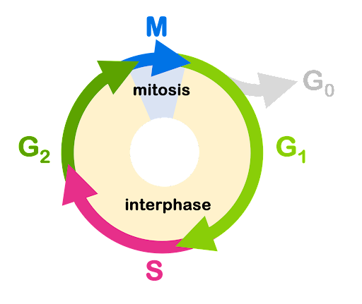
Although there is not much change visible under the light microscopy during interphase, in fact the cell performs a carefully orchestrated series of tasks during this time. Interphase is divided further into the stages of G1, S, and G2. This is illustrated in Figure 2.
Immediately following mitosis, the cells enter the G1 (Gap 1) phase of the cell cycle. During this phase, the cell performs normal metabolic functions and grows in size.
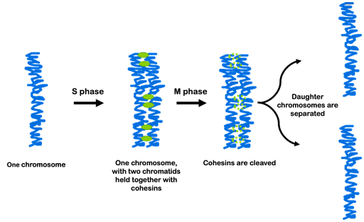
S phase is next. During S phase of the cell cycle, DNA Synthesis occurs. During this phase of the cell cycle, the cell must copy, or replicate, its entire genome in preparation for cell division. One complete copy must be available for each of the two daughter cells produced during cell division. The module on Replication describes this process more in detail. During S phase of the cell cycle, the DNA content of the cell doubles, with each chromosome now consisting of two chromatids. The chromatids remain connected via proteins called cohesins, and the total number of chromosomes does not change (Figure 3).
In G2 phase (Gap 2), the cell continues to grow and prepares for cell division. And in M phase (Mitosis), the cell carefully separates the two copies of the genome, partitioning them so the cell can divide into two daughter cells.
The progression from one stage to the next is driven by a series of cell cycle proteins called cyclins, and it is regulated by several checkpoints. At each checkpoint, the cell has mechanisms in place to ensure that conditions are right to move on to the next stage. For example, at the checkpoint between before the transition to S phase, the cell cycle will pause if there are insufficient cellular resources or DNA damage is detected. Replication of damaged DNA could have profound consequences for the cell and its daughters. If the cell proceeds through this checkpoint to S phase, the cell is now committed to cell division. At the G2/M checkpoint, the cell cycle will pause if DNA is incompletely replicated or if DNA damage is detected. If there are not two complete copies of the genome, this will impact the genomic integrity of daughter cells. There are additional checkpoints during M phase, which are discussed later in this module.
A cell can also exit the cell cycle and enter G0 phase from G1. Cells in G0 are described as quiescent. They still perform normal metabolic function, but they do not divide. Some cells can re-enter G1 phase in response to environmental triggers, but others can remain in G0 indefinitely: some eukaryotic cells like mature neurons or cardiac muscle cells may remain in G0 for the lifetime of an individual. This contributes to the difficulty in healing such tissues after injury: damaged cells cannot be regenerated or replenished if they are not dividing.
Test Your Understanding
Media Attributions
- The cell cycle © Amanda Simons is licensed under a CC BY-SA (Attribution ShareAlike) license
- DNA replication © Amanda Simons is licensed under a CC BY-SA (Attribution ShareAlike) license
- Uzbekov, R. & Prigent, C. A Journey through Time on the Discovery of Cell Cycle Regulation. Cells 11, 704 (2022). ↵
