DNA repair and cancer
Learning Objectives
- Define cancer, tumor, tumor-suppressor, proto-oncogene, oncogene.
- Compare and contrast mismatch repair, nucleotide excision repair, and base excision repair.
- Compare and contrast non-homologous end-joining and homologous recombination.
- Explain why cancer is described as a genetic disease.
- Describe the process by which a cell becomes cancerous, including the types of mutations in proto-oncogenes and tumor suppressors.
- Recognize the relationship between age and incidence of cancer.
- Explain why elephants and naked mole rates rarely get cancer, using the hallmarks of cancer to justify the explanation.
Source material:
Wong, EV. Cells – Molecules and Mechanisms1. Chapter 7.6 DNA Repair and Chapter 16: Viruses, Cancer, and the Immune System
https://bio.libretexts.org/Bookshelves/Cell_and_Molecular_Biology/Book%3A_Cells_-_Molecules_and_Mechanisms_(Wong)/16%3A_Viruses_Cancer_and_the_Immune_System/16.03%3A_Cancer [1]
Introduction
Before we get started, stop and think about what you know about cancer. You may think of words like “tumor.” You may have heard of people saying they “have the breast cancer gene.” You may have seen images of people who lose their hair as they undergo cancer treatments. In this chapter, we will look at the genetic mechanisms by which cancer arises.
The process by which cancer forms is called oncogenesis. The prefix “onco-” refers to cancer. You’ll see this used in words like “oncologist,” which is a doctor who specializes in cancer, or “oncogene,” which, as we will see, is a gene associated with oncogenesis.
A tumor is a mass of cells that grows much faster than surrounding healthy tissue. All multicellular organisms can grow tumors – even plants, as shown in Figure 1. A tumor typically arises from one cell that begins to proliferate faster than its surrounding cells.

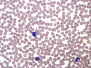
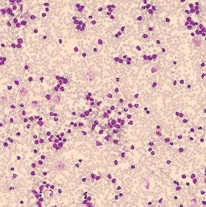
Tumors can be a solid mass of cells like those seen in Figure 1, or they can be “liquid tumors” – individual cells that are dividing uncontrollably but are not all clumped together. Blood cancers like leukemias are liquid tumors because there is no solid mass of cells, but the cancer cells end up overrunning the bloodstream. Leukemia is shown in Figure 2. On the left is a normal blood smear, where you can see many pink-red blood cells and far fewer purple-staining white blood cells. On the right is a blood smear from a leukemia patient. Note the abundance of purple-stained leukemia cells among red blood cells in the leukemia smear.


Test Your Understanding
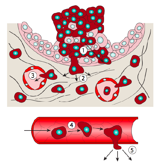
Tumors begin to cause health problems when they impede the normal function of the tissue in which they are found. For example, lung cancers will impede the function of the lungs and eventually block the ability to breathe. Tumors that arise in nonessential parts of the body – like breast tissue, for example – cause health problems not because they block the function of their tissue of origin but because they can metastasize. Metastasis means that cells escape from the original (or primary) tumor, travel through the bloodstream, and form a secondary tumor at a distant site. The process of metastasis is described in more detail in Figure 3.

A tumor is not necessarily cancerous. A benign tumor stays localized to its original location in the body, surrounded by an extracellular matrix that encapsulates the tumor. A benign tumor can still cause health problems by putting pressure on surrounding healthy tissue and impeding normal function. However, a cancerous or malignant tumor has the potential to spread beyond its original boundaries and can metastasize to new tissue, impeding the function of both the primary tissue and the secondary tissue.
Cancers are usually defined by their tissue of origin. So, in humans, breast cancer arises because of the uncontrolled growth of breast cells, lung cancers are an uncontrolled growth of cells in the lung, and leukemias (a blood cancer) are the uncontrolled growth of leukocytes (a type of white blood cell). Even after metastasis, such cancers are still describable by their primary tissue.
The growth advantage that tumor cells have over their surroundings comes from the somatic mutation of genes involved in cell growth and maintenance of the genome. For this reason, cancer is said to be a genetic disease. You’ll recall from our discussion of mutation that somatic cells are not reproductive cells, so the cancer-associated somatic mutations will not be inherited by offspring.
In the chapter on mutations, we discussed some of ways mutations can arise. But DNA is damaged far more often than it is mutated because the vast majority of damage is repaired. Both prokaryotic and eukaryotic cells have specific mechanistic pathways to repair damage. These pathways are overlapping in function and some proteins are shared between pathways. When these repair pathways fail, mutations can accumulate. A mutation in exactly the wrong place of the genome can cause the cell proliferation that leads to the growth of a tumor.
This chapter will begin with a look at how damage arises and how different types of damage are repaired. We’ll then transition to the types of mutations that can lead to cancer, with some examples of specific genes involved in oncogenesis. Finally, we’ll talk briefly about cancer treatments, including a few examples of targeted therapies that exploit what we know about cancer genetics.
Test Your Understanding
Media Attributions
- Figure 1 DNA Repair is licensed under a CC0 (Creative Commons Zero) license
- Figure 2-left DNA Repair © By Keith Chambers - Scooter Project is licensed under a CC BY-SA (Attribution ShareAlike) license
- Figure 2-right DNA Repair © By El*Falaf is licensed under a CC BY-SA (Attribution ShareAlike) license
- Figure 3 DNA Repair © Metastasis is shared under a not declared license and was authored, remixed, and/or curated by LibreTexts. is licensed under a CC0 (Creative Commons Zero) license
- Cells - Molecules and Mechanisms (Wong). Biology LibreTexts https://bio.libretexts.org/Bookshelves/Cell_and_Molecular_Biology/Book%3A_Cells_-_Molecules_and_Mechanisms_(Wong) (2018). ↵

