Eukaryotic Gene Regulation in Action: Examples from Development
Learning Objectives
- Define homeotic mutation, differentiation, enhancer, enhanceosome, silencer, insulator, TAD, maternal effect gene, Hox gene.
- Explain how multiple enhancers can work to generate complex patterns of gene expression, using eve and Shh regulation as examples.
- Describe mechanisms by which chromatin structure can influence gene expression
- Describe the difference between a forward and reverse genetic screen.
- Describe how maternal effect, gap, pair-rule, segment polarity, and segment identity genes interact to direct body patterns in the developing embryo.
- Recognize that proper regulation of a gene doesn’t just mean the gene is turned on! It means that it is only turned on under proper conditions.
- Recognize that mutations in non-coding regulatory sequences can have profound effects on phenotype.
Introduction: How does a body get built? Homeotic mutations rearrange body structures
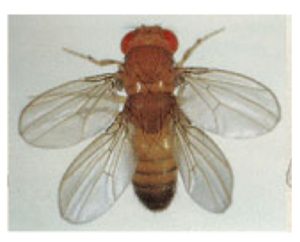
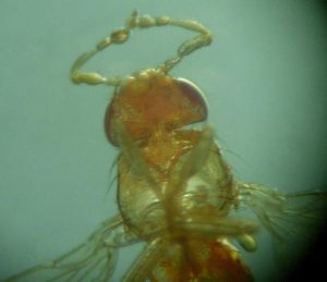
In the 1970’s, researchers like Edward Lewis were interested in mutations that resulted in a rearrangement of body structures in Drosophila, where one body part was replaced with another. The mutated genes were called homeotic genes. One such example was a mutation in the gene ultrabithorax (Ubx). Ubx mutants had an extra set of wings in place of the halteres, which in wild type flies are short structures important for stability (Figure 1a). Other mutants were missing expected body parts in particular segments. One mutant even had legs growing where antennae should be (Figure 1b)!


Around the same time, geneticists Eric Wieschaus and Christiane Nüsslein-Volhard were beginning to look at developmental mutant phenotypes, too. However, they were not interested in mutant phenotypes in the adult fly. They reasoned that the genes important for body patterning were likely to be so important that mutations would be embryonically lethal. So, instead, Wieschaus and Nüsslein-Volhard looked for mutant phenotypes in the Drosophila embryo.
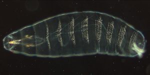
About 24 hours after fertilization of the egg, a wild-type Drosophila embryo looks like Figure 2, with visible segments. In this image, the anterior (head) part of the embryo is to the left, and the posterior (tail) is to the right. You can watch a video of the early stages of Drosophila development on YouTube here and here too. You can see that each segment is slightly different from the others. As development progresses, the segments each eventually assume a different identity. For example, one segment will produce antennae, one will produce a wing, etc.

Figure 2. Drosophila embryos have a segmented appearance. The light-colored spots are denticles, short projections that extend from the ventral side of the embryo. Each segment has a slightly different denticle pattern.
How does a single-cell zygote develop into a multicellular organism, with specialized cells, distinct tissues, and organ systems? And how do those processes go wrong in the homeotic mutants? The differences in cell and tissue types largely come down to differences in gene regulation.
In this module, we look at how genes are regulated in eukaryotes, using embryonic development in animals as an example. We will review the basics of transcription and gene regulation, and then dive in to see how gene regulation drives cell differentiation during embryonic development. We’ll look at how multiple enhancer elements can regulate gene function, using the Drosophila gene eve as an example.
Media Attributions
- Figure 1a EG Regulation © Matthew Slattery, et al is licensed under a CC BY-SA (Attribution ShareAlike) license
- Figure1b EG Regulation © File: Mutation_Antennapedia.jpg is licensed under a Public Domain license
- Figure 2 EG Regulation is licensed under a Public Domain license

